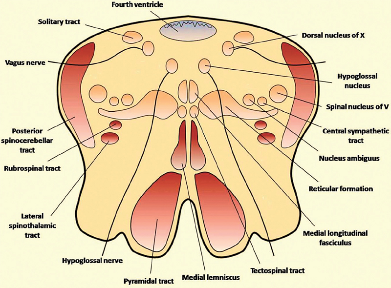
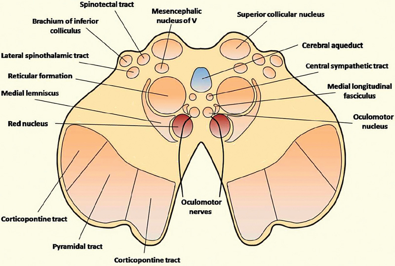
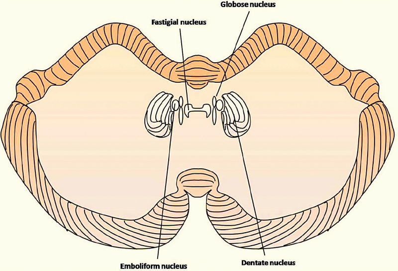
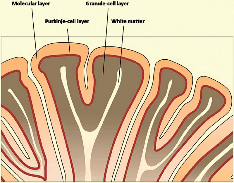
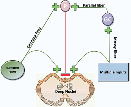
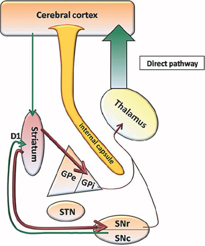
16 What are the three brainstem segments (from caudal to rostral)? Medulla, pons, midbrain Into what three segments are the medulla, pons, and midbrain further divided? Tectum (“roof,” dorsal aspect), tegmentum (ventral aspect), basis (most ventral aspect of tegmentum) Which artery or arteries supply the medulla? Branches of the vertebral arteries, posterior inferior cerebellar artery (PICA), anterior spinal artery, and posterior spinal artery The presence or absence of what structure designates the rostral and caudal portions of the medulla, respectively? Fourth ventricle (present in rostral medulla, absent in caudal medulla)1 Which ascending pathways are present in the medulla? Dorsal columnar (medial lemniscal), spinothalamic, spinoreticular, and spinocerebellar Which descending pathways are present in the medulla, and what functions do they mediate? Corticospinal tract (motor), descending spinal tract of CN V, medial longitudinal fasciculus (gaze and head movements), and tectospinal tract (neck and trunk movements in response to visual stimuli) What fibers do the inferior cerebellar peduncles contain? Fibers efferent from spinal cord and medulla to cerebellum, and crossed olivocerebellar fibers Which cranial nerve nuclei are housed within the medulla, and what functions do they mediate? Hypoglossal nucleus (motor to tongue), dorsal motor nucleus of X (parasympathetic innervation of viscera), and solitary tract and nucleus (taste and visceral sensory to ventral posteromedial nucleus [VPM] of thalamus) Which cranial nerves exit the brainstem via the medulla, and through which foramina do they exit the skull? VII and VIII exit via the internal acoustic meatus of the petrous temporal bone in the posterior fossa. IX and X emerge from a shallow groove dorsal to the olive, IX to XI exit via the jugular foramen, and XII emerges from the anterolateral sulcus and exits through the hypoglossal foramina of the occipital bone. Fig. 16.1 Cross-sectional illustration of rostral medullary anatomy. How is the homunculus organized in the medullary medial lemniscus? Cervical sensory information is dorsal/posterior to sacral information. Occlusion of what artery or arteries leads to medial medullary syndrome? Vertebral branches or the anterior spinal artery What are the symptoms of medial medullary syndrome? • Contralateral hemiparesis (due to damage to the ipsilateral pyramid) • Contralateral sensory deficits (due to damage to the medial lemniscus) • Ipsilateral paralysis and atrophy of tongue muscles (due to damage to the hypoglossal nucleus and/or nerve) Occlusion of what artery or arteries lead to lateral medullary (aka Wallenberg’s) syndrome? Vertebral branches (most commonly)2 or PICA What are the symptoms of Wallenberg’s syndrome?2 • Loss of pain and temperature sensation over contralateral body (due to damage to the spinothalamic tract) with relative sparing of tactile sensation • Loss of pain and temperature sensation over ipsilateral face (due to damage to the spinal trigeminal tract) • Hoarseness and dysphagia (due to damage to the nucleus ambiguous) • Ipsilateral Horner’s syndrome (due to damage to the descending sympathetic fibers) • Possible vertigo, abnormal eye movements and ipsilateral cerebellar deficits (due to damage to the inferior cerebellar peduncle and adjacent vestibular nuclei) Occlusion of what artery or arteries lead to Weber’s syndrome? Branch of the posterior cerebral artery What gross structure is damaged in Weber’s syndrome? Cerebral peduncle What are the symptoms of Weber’s syndrome? • Contralateral spastic paresis (due to damage to corticospinal fibers) • Ipsilateral ptosis, pupillary dilation, and lateral strabismus (due to damage to the occulomotor nerve) Which artery or arteries supply the pons? Basilar artery and its branches (paramedian and circumferential branches), with some contribution from the anterior inferior cerebellar artery (AICA) and superior cerebellar arteries Which structures are contained within the basis pontis? Corticospinal fibers, pontine nuclei (contain motor information derived from the cerebral cortex via the corticopontine pathway), and pontocerebellar fibers (relay information from pontine nuclei to cerebellum via the middle cerebellar peduncle Which neurotransmitter is contained within the raphe nuclei? Serotonin (the raphe nuclei mediate arousal, the sleep-wake cycle, and sensation of painful stimuli) Which cranial nerve nuclei are contained within the pontine tegmentum? Main sensory nucleus of V, the abducens nucleus (VI), and the facial nucleus (VII) Which fibers are contained within the central tegmental tract? Descending fibers from the midbrain to the inferior olivary nucleus, and ascending fibers from the reticular formation to the thalamus What course does the abducens nerve (VI) follow after exiting the pons? Travels medially in the middle cranial fossa, enters the carotid sinus, and exits the skull via the superior orbital fissure along with V1 (ophthalmic branch of trigeminal nerve) How is the homunculus oriented in the pontine medial lemniscus? The feet are represented laterally, with the cervical sensory input represented medially. Which artery or arteries supply the midbrain with blood? Posterior cerebral arteries with some contribution from the basilar artery branches and the superior cerebellar arteries Which three important structures are contained within the base of the midbrain? Crus cerebri (includes corticospinal, corticobulbar, and corticopontine fibers), substantia nigra (a mediator of motor control), and cerebral peduncle To what structure does the substantia nigra send efferent dopaminergic fibers? Striatum What fibers are contained within the cerebral peduncle? Descending corticospinal and corticopontine fibers Which cranial nerve nuclei lie within the midbrain? Trochlear nucleus (IV) and occulomotor nuclei (III) Which neurotransmitter is contained within the locus ceruleus nucleus? Norepinephrine (the locus ceruleus regulates arousal and the sleep-wake cycle) What sort of information is received and processed by the superior and inferior colliculi? Visual and auditory information, respectively Which fibers are contained within the superior cerebellar peduncle? Fibers efferent from the cerebellum to the red nucleus and spinocerebellar tracts Where do the occulomotor nerves exit the midbrain? Ventrally/anteriorly, between the cerebral peduncles in the interpeduncular fossa Fig. 16.2 Cross-sectional illustration of midbrain anatomy. What is the basic gross organization of the cerebellum? Two lateral cerebellar hemispheres surround a midline vermis. A cerebellar cortex overlies the cerebellar white matter, where four paired deep nuclei are located posterior to the fourth ventricle. What are the names of the four deep cerebellar nuclei (from medial to lateral)? Fastigial nuclei, globose nuclei, emboliform nuclei, dentate nuclei Fig. 16.3 Deep cerebellar nuclei. What is the role of the cerebellar vermis? Axial control (controls muscle tone and coordination of the trunk) What is the role of the cerebellar hemispheres? Each controls tone and coordination of the ipsilateral appendages. What are the names of the phylogenetic segments of the cerebellum and what are their roles? Archicerebellum (contains flocculus and nodulus, and plays a role in posture, balance, eye movements); paleocerebellum (plays a role in lower limb movements and speech); and neocerebellum (plays a role in limb coordination) Multiple synaptic contacts between mossy fibers with Golgi and granule cells that occur at the granular layer; Glomeruli = Golgi and Granule What are the three cytoarchitectural layers of the cerebellar cortex (from superficial to deep)? An outer molecular layer, a Purkinje-cell layer, and an inner granule cell layer3 Fig. 16.4 Cytoarchitectural organization of cerebellar cortex. Where do climbing fibers originate? In the contralateral inferior olivary nucleus What neurotransmitter is used by the climbing fibers? Aspartate Where do mossy fibers originate? Deep cerebellar nuclei, vestibular nuclei, the spinal cord, and the cerebellar cortex (via pontine nuclei) Fig. 16.5 Schematic diagram of inputs to deep cerebellar nuclei. P, Purkinje cells; GC, granule cells; (+), excitatory; (−), inhibitory. Which peduncle is primarily efferent? The superior cerebellar peduncle Where does the superior cerebellar peduncle attach to the brainstem? Dorsally in the rostral pons, near the pontinemidbrain junction Which cranial nerve emerges near the superior cerebellar peduncle? Trochlear (CN IV) Which fibers travel through the superior cerebellar peduncle? Input: fibers from the contralateral spinal cord terminating in the ipsilateral vermis and intermediate zone (anterior spinocerebellar tract) Which peduncle contains only afferent input? The middle peduncle Where does the middle cerebellar peduncle attach to the brainstem? The caudal pons Which fibers travel through the middle cerebellar peduncle? Input: fibers from cerebral cortex (via pontine nuclei) terminating in the contralateral cerebellum (pontocerebellar tract) Where does the inferior cerebellar peduncle attach to the brainstem? Dorsolaterally in the rostral medulla Which fibers travel through the inferior cerebellar peduncle? Input: fibers from Clarke’s nucleus terminating in the ipsilateral vermis and intermediate zone (posterior spinocerebellar tract), fibers from the lateral cuneate nucleus terminating in the ipsilateral vermis and intermediate zone (cuneocerebellar tract), fibers from the vestibular ganglion and vestibular nuclei terminating in the vermis (vestibulocerebellar tract), fibers from the main sensory and spinal nuclei of V terminating in the ipsilateral vermis and intermediate zone (trigeminocerebellar tract), fibers from the inferior olivary nucleus terminating in the contralateral cerebellar cortex (olivocerebellar tract) Output: fibers from the deep cerebellar nuclei terminating in the contralateral red nucleus or contralateral thalamic motor nuclei (ventral lateral and ventral anterior nuclei) What are the alternate names for the cerebellar peduncles? Superior is the brachium conjunctivum Middle is the brachium pontis Inferior is the corpus restiform or restiform body Damage to the midline cerebellum causes what deficit? Postural instability Damage to the lateral cerebellum causes what deficit? Limb ataxia Damage to the flocculus causes what deficit? Difficulty with eye movements (poor pursuit and voluntary eye movements) The thalamus is divided into three major nuclear masses (anterior, medial, and lateral) by what white matter bundle? Internal medullary lamina What is the role of the thalamus? Serves as a primary relay center for all information passing from the brainstem and spinal cord into the cerebral cortex and also receives reciprocal projections from the cerebral cortex. What are the boundaries of the thalamus? Posterior limb of the internal capsule laterally, the third ventricle medially, the subthalamus and hypothalamus inferiorly, and the body of the lateral ventricle and corpus callosum superiorly In which portion of the hypothalamus does the lamina terminalis lie? The anterior portion (the chiasmatic region) What structures are contained within the central portion? The tuber cinereum and infundibulum What structures are contained within the posterior portion? Mammillary bodies Which hypothalamic nuclei release peptide hormones from the posterior pituitary or neurohypophysis? Supraoptic and paraventricular nuclei Which two peptide hormones are released from the posterior pituitary? Antidiuretic hormone (vasopressin) and oxytocin Which structure within the subthalamus comprises a portion of the basal ganglia? Subthalamic nucleus (body of Luys) From what structure does the subthalamic nucleus receive input? Globus pallidus To what structure do fibers from the subthalamic nucleus project? Globus pallidus What neurological symptom would one expect from lesions of the subthalamic nucleus? Contralateral hemiballismus Which midline structure is contained within the epithalamus? Pineal gland Which cerebral cortex lies deep to the sylvian fissure? Insular cortex Which fissure/sulcus contains the primary visual cortex? Calcarine fissure Which sulcus forms the anterior boundary for the occipital lobe? Occipitoparietal sulcus What are the names of the two gyri forming the medial surface of the parietal lobe? Posterior paracentral lobule and precuneus What are the names of the three gyri forming the medial surface of the occipital lobe? From superior to inferior: cuneate gyrus, lingual gyrus, occipitotemporal or fusiform gyrus With what gyrus is the lingual gyrus continuous anteriorly? Parahippocampal gyrus What structure marks the transition from lingual to parahippocampal gyri? The isthmus of the cingulate gyrus Where does Broca’s area reside? The opercular and triangular portions of the inferior frontal gyrus of one hemisphere (typically the left) What neurological deficit would one expect with a lesion to Broca’s area? Expressive or “nonfluent” aphasia Where does Wernicke’s area reside? The posterior portion of the superior temporal gyrus of one hemisphere (typically the left) What neurological deficit would one expect with a lesion to Wernicke’s area? Receptive or “fluent” aphasia What is a pyramidal cell? The most numerous neurons in the neocortex, they consist of a cone-shaped cell body, a single, long apical dendrite projecting toward the cortical surface, and several basal dendrites spreading outwardly from the base of the cell body. What are dendritic spines? Small, membranous projections from dendrites containing synaptic contacts What are the six layers of the neocortex? From superficial to deep: 1. Molecular layer 2. External granular layer 3. External pyramidal layer 4. Internal granular layer 5. Internal pyramidal layer 6. Multiform layer Which gyri/areas comprise the limbic lobe? Cingulate and parahippocampal gyri as well as the subcallosal area (inferior to the genu of the corpus callosum) and the paraterminal gyrus (anterior to the lamina terminalis); together with the hippocampus and amygdala, these structures comprise the limbic system. What is the primary role of the limbic system? Control of basic survival functions (the four F’s: feeding, fighting, fleeing, fornicating) Which structure forms the primary output pathway for the hippocampus? The fornix From what area do the majority of hippocampal inputs arise? Entorhinal cortex (which receives inputs from the olfactory cortex, cingulate gyrus, orbital cortex, amygdala and other temporal lobe areas) What is the role of the basal ganglia? Stores learned movements, and controls intensity and timing of movements What are the five major nuclei forming the basal ganglia? Caudate and putamen (together known as the striatum), globus pallidus, subthalamic nuclei, and the substantia nigra Which nuclei are collectively known as the “lentiform nucleus”? Putamen and globus pallidus Excitatory influences in the basal ganglia are mediated by which neurotransmitter? Glutamate Inhibitory influences in the basal ganglia are mediated by which neurotransmitter? GABA From where does the primary input to the basal ganglia arise? The cerebral cortex (primarily motor and premotor areas) projects via the internal and external capsules to the striatum, where it exerts an excitatory effect. What other structures form inputs to the striatum? The substantia nigra pars compacta forms divergent inputs to the striatum (the nigrostriatal pathway), those with an excitatory influence (mediated by the D1 receptor) and those with an inhibitory (mediated by the D2 receptor) influence on the striatum From where do the primary outputs from the basal ganglia arise? The primary basal ganglia outputs arise in the globus pallidus and substantia nigra pars reticulata. Describe the direct pathway. The cerebral cortex exerts an excitatory influence on the striatum, which in turn exerts an inhibitory influence on both the globus pallidus internal segment (GPi) and substantia nigra pars reticulata (SNr). Together, the GPi and SNr inhibit the thalamus, which in turn excites the cerebral cortex. What is the net effect of the direct pathway on the thalamus and cerebral cortex? Because the pathway results in increased inhibition of the GPi and SNr (which normally inhibit the thalamus), the pathway causes a net excitation (mediated by glutamate) of the thalamus and motor cortex, stimulating movement. Inhibition of an inhibiting agent, as in this case, is known as disinhibition and results in net activation. Fig. 16.6 Basal ganglia direct pathway. D, dopamine; GPe, external globus pallidus; GPi, internal globus pallidus; SNc, substantia nigra pars compacta; SNr, substantia nigra pars reticularis; STN, subthalamic nuclei.
Neuroanatomy
16.1 Brainstem and Cranial Nerves
16.1.1 Medulla
16.1.2 Pons
16.1.3 Midbrain
16.2 Cerebellum
16.3 Diencephalon
16.3.1 Thalamus
16.3.2 Hypothalamus
16.3.3 Subthalamus
16.3.4 Epithalamus
16.4 Cerebral Hemispheres/Telencephalon
16.4.1 Limbic System
16.4.2 Basal Ganglia
![]()
Stay updated, free articles. Join our Telegram channel


Full access? Get Clinical Tree


