Neurologic Critical Care


Neurologic Critical Care


Réjean M. Guerriero

Patricia L. Musolino

Robert C. Tasker


INTRODUCTION

The acute neurologic care of critically ill patients and a critical care approach to acute neurologic conditions are emerging interests within the field of pediatric neurology. The medical conditions discussed in this chapter are neurologic emergencies.


AN APPROACH TO THE NEUROLOGICALLY CRITICALLY ILL CHILD

To approach a patient with a disorder of consciousness requires rapid directed review of the history and a focused neurologic exam.1

HISTORY: Should be acquired from relatives and witnesses and should include:


  • Onset: acute, subacute, and insidious


  • Preceding symptoms: headache, vertigo, vision or hearing complaints, focal neurologic findings, fever, and signs of systemic illness


  • Environment: recent trauma, access to toxins or medications


  • Medical history: medical problems (epilepsy), medications

GENERAL PHYSICAL EXAM: Focused on vital signs, respiratory pattern, and hemodynamics; also observe for evidence of trauma, infection, nuchal rigidity.

NEUROLOGIC EXAM: Should focus on mental status, level of brainstem dysfunction, and focal neurologic findings. Coma scales may be useful, but a descriptive exam is generally preferred.


  • Mental Status: Eyes open or closed? Responds or regards? Follows any commands?


  • Cranial Nerves: Blink to threat (CN II). Reactivity of pupils (CN III). Spontaneous eye movements or tracking (CN III, IV, VI). Corneal reflexes (CN V, VII). Oculocephalic reflex, assuming no cervical trauma (CN VIII, III, VI). Gag (CN IX, X, XII).


  • Motor: Asymmetries in tone or spontaneous movements. Response to noxious stimuli with localizing, withdrawal, or posturing. Asymmetries in reflexes.

DIFFERENTIAL DIAGNOSIS: The differential diagnosis for altered consciousness is broad. Many of the possible diagnoses, including trauma, anoxia, seizures, infection, and metabolic encephalopathy, are covered in this chapter.

ICU MONITORING: There are various neuroimaging techniques, as well as invasive monitoring, that may be useful. See Table 13.1.


ACUTE INTRACRANIAL HYPERTENSION

Assessing the degree of brain oxygenation is a fundamental step in the care of neurologic emergencies. In this section we will first address the normal
physiology of cerebral perfusion and brain oxygenation, followed by general pathophysiology.


TABLE 13.1 Neuromonitoring in the ICU


Technique


Benefits/Utility


Clinical Scenario


Computed tomography (CT)


Quick, low cost. Negative = radiation exposure


Hemorrhage, infarction, mass lesion, bony lesion


Head ultrasound (HUS)


Noninvasive, lower cost, no radiation. Requires an open fontanel


IVH, larger hemorrhages, and mass effect, serial monitoring


MRI


Conventional imaging (T1, T2)


Structure, CSF


Structural lesion, edema


Diffusion-weighted imaging (DWI) and apparent diffusion coefficient (ADC)


Areas of restricted diffusion


Hypoxic and/or ischemic injury, abscess


T1 with gadolinium contrast


Identifies areas of blood-brain barrier breakdown


Tumor, abscess


Magnetic resonance angiography (MRA)


Vessel integrity and path


Thrombosis, dissection, vascular malformations


Magnetic resonance venography (MRV)


Venous integrity and path


Thrombosis, vascular malformations


Arterial spin labeling


Perfusion study without radiation


Areas of decreased (or increased) perfusion


Susceptibility-weighted imaging (SWI) or gradient echo (GRE)


Assessing venous blood and iron


Microhemorrhages, contusion, thrombosis


Magenetic resonance spectroscopy (MRS)


Assesses for metabolic abnormalities


Metabolic encephalopathy, trauma, hypoxic-ischemic injury


Bedside monitoring


EEG


Continuous, real-time assessment of cortical activity


Seizure detection, burst suppression, encephalopathy


Near infrared spectroscopy (NIRS)


Continuous, real-time brain oxygenation assessment (Arterial – Venous O2)


Coma, encephalopathy, trauma


Transcranial Doppler


Noninvasive


Vasospasm, perfusion


Intracranial pressure (ICP) monitoring


Direct measurement of ICP. Negative = invasive


Any condition with concerns for increased ICP


Note: See Chapter 3 for more details.


NORMAL PHYSIOLOGY: The brain requires oxygen and glucose for its metabolic needs, which requires maintaining cerebral blood flow (CBF).


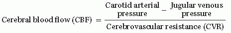
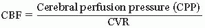
  • CPP = Mean arterial pressure (MAP) – Intracranial pressure (ICP)


    • MAP = 2/3 Diastolic pressure (DP) + 1/3 Systolic pressure (SP)


    • MAP = DP + 1/3 (SP – DP)

      Therefore, increases in blood pressure lead to increases in CPP and increases in ICP lead to decreases in CPP


  • Normal range for CPP is age dependent since MAP increases with age while ICP remains mostly unchanged (Table 13.2; note that these values vary slightly based on gender and height)


  • Cerebral vasculature mechanisms to maintain CPP (Fig. 13.1)


    • Metabolic mechanisms based on plasma osmolality


    • Neural mechanisms can be intrinsic and extrinsic


      • Intrinsic: Autoregulation (A, Fig. 13.1)


      • Extrinsic: PaCO2: 35-45 mm Hg, increased PaCO2 (B) leads to vasodilation, which increases CBF and indirectly the ICP (C). Conversely, decreasing the PaCO2 with hyperventilation will acutely lower the ICP. Finally, when the pO2 is low (<50 mmHg), CBF increases, as can the ICP (D).

ETIOLOGY: Brain edema from traumatic brain injury (TBI), hypoxic-ischemic encephalopathy (HIE), or ischemic stroke as well as bleeding or expanding lesion are common causes. In infants and children this may also
occur with dysfunctions of ventricular shunts and their valves (ventriculosubgaleal, ventriculoperitoneal, ventriculoatrial, and ventriculopleural shunts).


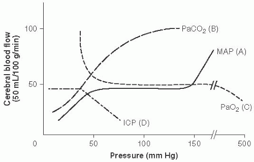
FIGURE 13.1 Cerebral Vasculature Mechanisms to Maintain Cerebral Perfusion Pressure (CPP). Cerebral blood flow (CBF) = CPP over cerebral vascular resistance (CVR). With a MAP 50 to 150 mm Hg in older children and adults (lower range in infants and younger children). Autoregulation maintains stable CBF with changes in CVR. (Adapted with permission from Westover MB, Choi E, Awad KM, et al., eds. Pocket Neurology. Philadelphia, PA: Lippincott, Williams and Wilkins; 2010:46-47.)

General Pathophysiology:


  • The cranial vault contains:


    • Brain parenchyma, 80%; blood, 10%; CSF, 10%.


    • The amount of CSF ˜150 mL, with 500 mL made daily (about 20 mL/h).


  • In adults and older children the ICP is generally between 11 and 28 cm H2O or 8 to 20 mm Hg, although this range decreases with age2 (see Table 13.2).


  • Any increase in brain parenchyma, blood, or CSF can produce changes in mechanical forces and may lead to herniation (Fig. 13.2).


  • Both cytotoxic and vasogenic mechanisms can lead to cerebral edema.


    • Cytotoxic edema is caused by cellular injury that leads to excitatory amino acid release, leading to cell depolarization and sodium traveling into the cell, causing swelling.


    • Vasogenic edema is the result of dysfunctional vessels, such as those of a tumor, which allow passage of intravascular fluid into the interstitial space.

CLINICAL FEATURES: Symptoms include headache, vomiting, vision changes, and gaze palsies. Impending herniation may be heralded by Cushing’s triad, which includes hypertension, bradycardia, and irregular respirations.


TRAUMATIC BRAIN INJURY

DEFINITION: TBI is one of the leading causes of trauma-related hospitalizations and mortality in children. It occurs when an external mechanical force causes brain dysfunction.

ETIOLOGY: In children the causes vary by age, with falls being the leading cause in children 0 to 4 yo and motor vehicle accidents being by far the leading cause in adolescence, ages 15 to 19. Nonaccidental head trauma in young children is an important cause of TBI and should always be assessed for. It is the leading cause of abuse-related deaths in the United States.3

EPIDEMIOLOGY: Recent estimates by the CDC reveal that from 2002 to 2006 there were 511,257 traumatic brain injuries in children 0 to 14 yo that led to 473,947 ED visits, 35,136 hospitalizations, and 2,174 deaths annually.3

PATHOPHYSIOLOGY: The biomechanics of TBI involve both linear and/or rotation forces. Linear forces, such as those resulting from a fall, tend
to cause brain contusions, focal injuries, and coup-contrecoup injuries. Rotational injuries occur in motor vehicle accidents and sports injuries, and nonaccidental trauma leads to shearing forces causing traumatic axonal injury occurring in white matter fiber tracts. All of these injuries lead to increased excitatory neurotransmission and neurometabolic cascades, leading to axonal injury and cell death.4 Furthermore, children’s brains seem to be more susceptible to cerebral swelling following head injury as compared with those of adults.5


TABLE 13.2 Normal Heart and Respiratory Rates, Blood Pressure, Mean Arterial Pressure (MAP), Intracranial Pressure (ICP), and Cerebral Perfusion Pressure (CPP) for Different Ages2,55,56,57


Age


Heart Rate (beats/ min)


Resp. Rate (breaths/ min)


Blood Pressure (mm Hg)


MAP (mm Hg)


ICP (mm Hg)


CPP (mm Hg)


0-3 mo


100-150


35-55


65-85/45-55


35-65


2-5 (3-7 cm H2O)


>30


3-12 mo


80-120


25-45


70-100/50-65


55-75


1-6 y


65-110


20-30


85-110/45-75


65-85


3-7 (4-10 cm H2O)


>40


6-12 y


60-95


14-22


95-120/55-80


70-90


>50


>12 y


60-100


12-18


100-130/60-80


70-95


< 20 (11-28 cm H2O)


>60


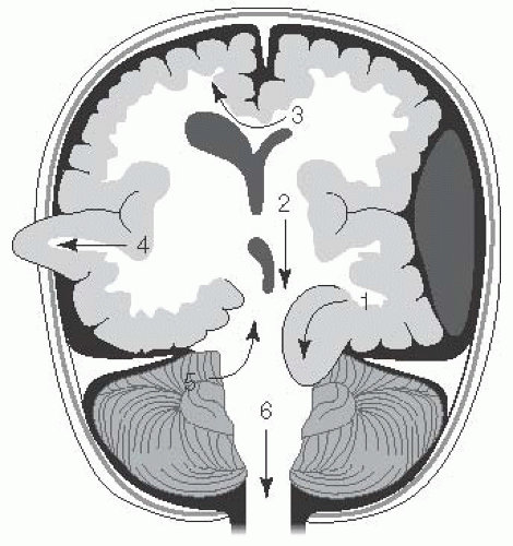
FIGURE 13.2 Types of Brain Herniation. 1. Uncal: ipsilateral CN III palsy and contralateral hemiplegia or posturing. 2. Central transtentorial: coma with bilateral small pupils and progression of posturing from decorticate to decerebrate with loss of brainstem reflexes. 3. Subfalcine: coma and contralateral weakness to posturing. 4. Extracranial (when craniectomy is present): deficits from herniated territory. 5. Upward cerebellar: cerebellar symptoms to coma and bilateral posturing. 6. Tonsillar (downward cerebellar): decreased arousal, pyramidal signs, respiratory insufficiency, coma. (Adapted with permission from Westover MB, Choi E, Awad KM, et al., eds. Pocket Neurology. Philadelphia, PA: Lippincott Williams and Wilkins; 2010:46-47.)

CLINICAL FEATURES: The history and exam should be focused as discussed earlier. Mechanism of injury and the environment in which the injury occurred (e.g., seat belt, fall from height, drugs or alcohol involved) will guide diagnosis and treatment. One should assess for fractures by periorbital or retroauricular ecchymoses (“raccoon eyes” or “Battle’s sign”), an asymmetric fixed pupil, neck trauma, or other signs suggesting the need for emergent neurosurgical intervention.

DIAGNOSTIC WORKUP:


  • Noncontrast head CT remains the best neuroimaging modality for quickly assessing for bony abnormalities, hemorrhage, and swelling.


  • MRI will be more sensitive for ischemia and subtler structural abnormalities.


  • Lab testing should be focused on assessing for cause of the injury (urine toxicology) and systemic injury (e.g., liver and renal function).


MANAGEMENT: Includes both medical and neurosurgical interventions aimed at addressing cerebral swelling and neuroprotection (2012, updated guidelines).6


  • Osm 300-360


  • Measure BP (MAP). Goals: Maintain CPP (see Table 13.2)


  • PaCO2 35-40

MONITORING: ICP monitoring or EEG may be indicated when neurologic examination is limited by impaired consciousness or paralysis. Dysautonomia occurs in 10% of children following TBI and should also be carefully monitored for.7

TREATMENTS: Includes osmotherapy, sedation, and supportive measures8 (Table 13.3).

PROGNOSIS: Mortality from severe traumatic brain injuries in patients admitted to the PICU ranges from 9% to 22%.9,10 About a 3rd of patients will have severe disability, and 40% to 50% will have moderate disability, with only 11% to 17% having good recovery. All of these numbers appear to be improved when a pediatric neurocritical care program is instituted.11 Patients with TBI may be left with cognitive and/or motor deficits. The degree of traumatic axonal injury and global white matter pathology is likely related to the degree of cognitive deficits.12


HYPOXIC-ISCHEMIC ENCEPHALOPATHY

HIE in the pediatric population most commonly happens in the perinatal period (see Chapter 19); however, a number of insults that lead to cardiac or respiratory arrest have implications in older pediatric patients as well.

DEFINITION: HIE is the loss of oxygen or blood flow to the brain, leading to brain damage and neurologic sequelae.

ETIOLOGY: The etiology of HIE differs significantly between infant, children, and adult patient populations. In newborn infants HIE occurs in the perinatal period and may be related to hypotension, respiratory failure, infection, and can be related to metabolic or genetic factors. In adults hypoxic-ischemic injury is typically related to cardiac arrhythmia, including ventricular fibrillation or tachycardia. In the pediatric population, asphyxia, such as caused by drowning, upper or lower airway obstruction, sudden infant death syndrome (SIDS), trauma, and other causes leading to asystole or bradyarrhythmias are much more common.13

EPIDEMIOLOGY: 16,000 children die annually from cardiopulmonary arrest.14 The most common causes of noncardiac arrest include drowning, SIDS, trauma, intoxications, respiratory causes, such as asthma, aspiration, or other acute airway obstruction, and sepsis15; however, the rates of death from SIDS have been cut in half in the last 15 y.16 Cardiac etiologies are the most common cause of cardiopulmonary arrest in infants beyond the perinatal period.17 The most common causes of cardiac causes of arrest in patients 0 to 13 yo are congenital abnormalities, while primary arrhythmias were the most common in children 14 to 24 yo.18

PATHOPHYSIOLOGY: The brain requires large amounts of oxygen and glucose for cellular metabolic demands. When decreased blood oxygenation (hypoxia) or blood flow (ischemia) occurs, this leads to loss of energy reserves in the form of adenosine 5-triphosphate (ATP). ATP depletion maintains membrane gradients,19 and disruption of these begins a cascade of detrimental events, including cell depolarization, glutamate release, and
ultimately Ca2+ influx into the cells, leading to cell necrosis and apoptosis. Additionally, creation of free radicals, accumulation of toxic by-products, and local inflammatory response, as well as reperfusion injury contribute to cell necrosis and death.20


TABLE 13.3 Treatment of Increased Intracranial Pressure (ICP)


Goal


Treatment/Medication


Mechanism/Benefits/Risk


Osm therapies:


  • Osm 300-360


  • Maintain CPP


  • CPP = MAP – ICP


  • ICP < 20


  • CPP > 40


20% mannitol Bolus: 1 g/kg Maint: 0.25-0.5 g/kg q2-6h


1. Decreases blood viscosity increasing CPP (rapid, transient)


2. Osmotic effect (water from parenchyma drawn into vessel). Note: requires intact blood-brain barrier (BBB)


Risk: accumulation in areas of BBB breakdown


Hypertonic saline 3% acute use: 6.5-10 mL/kg


3% maintenance: 0.1-1 mL/kg/h


23% rarely used because of safety in small patients


1. Osmotic benefit (sodium does not cross the BBB well)


2. Theoretical: improved resting membrane potentials; decreased inflammation


Risk: rebound in ICP


PaCO2 35-40


Hyperventilation


Induces hypocarbia → vasoconstriction


Mild: PaCO2 30-35 mm Hg


Aggressive: can be considered as a temporizing measure with PaCO2<30 mm Hg in the first 48 h


Risk: iatrogenic ischemia


Decrease brain metabolism


Anesthetics e.g., barbiturates


1. Reduce oxygen demand


2. Decreased electrical activity


3. Decreased neurotransmitter synthesis


Quiet, normothermia (preventing hyperthermia)


Decreases brain excitation/activity, thereby decreasing oxygen demand


General measures


Pressors


Head of bed 30°


Maintain MAP Reduces ICP

Only gold members can continue reading. Log In or Register to continue

Stay updated, free articles. Join our Telegram channel

Jun 20, 2016 | Posted by in NEUROLOGY | Comments Off on Neurologic Critical Care

Full access? Get Clinical Tree

Get Clinical Tree app for offline access