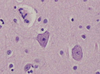
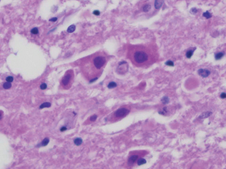
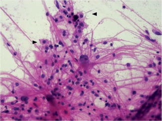
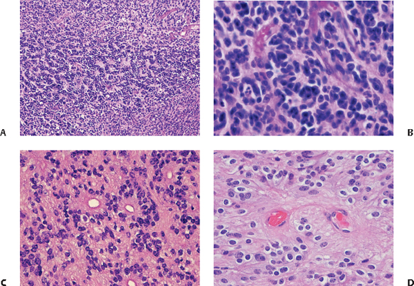
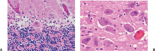
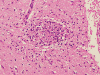
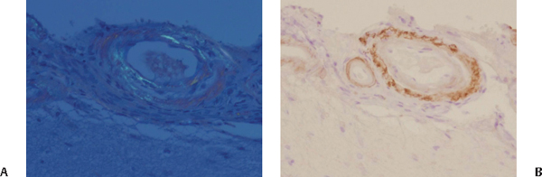
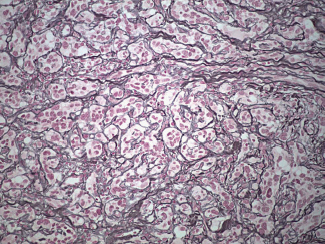
20 What features do neurons generally have in common? Round nucleus with prominent nucleolus and soma with lots of rough ER (Nissl substance)1–6 Fig. 20.1 Pyramidal neurons have round nuclei with a prominent nucleolus. The soma is pink-purple (amphophilic), due to abundant rough endoplasmic reticulum (Nissl substance). Motor neurons are trapezoidal, whereas sensory neurons are globular. The background neurophil is homogeneous, and the cell bodies of resting astrocytes cannot be seen. Which kinds of neurons are globular in shape, and which kinds are more trapezoidal? Sensory are globular, and motor are trapezoidal How are granular cell neurons different? They are much smaller and do not always show cell processes in normal sections. Where are they commonly found? Commonly found in the cerebellum and dentate gyrus of hippocampus What changes are indicative of irreversible neuron death? “Red is dead”: The neuron shrinks; the basophilic cytoplasm becomes eosinophilic; the nucleus becomes dark and shrunken, with loss of nuclear detail (pyknosis); occurs within 6 hours of ischemic necrosis.1–6 Fig. 20.2 Irreversibly damaged “red neurons” are shrunken. The normally basophilic cytoplasm becomes eosinophilic. The nucleus is pyknotic, with shrinking, hyperchromasia, and loss of nuclear detail. What is ferrugination? The coating of dead neurons with Fe2+ and Ca2+ salts Fig. 20.3 Dead, ferruginated neurons are coated with Fe2+ and Ca2+ salts. Give three examples of progressive or reversible neuronal injury. 1. Central chromatolysis 2. Neurofibrillary tangles 3. Neuronal storage of lipid or carbohydrates When does central chromatolysis occur? After axonal injury What changes are seen histologically? Nissl substance disappears, soma enlarges, and the nucleus becomes eccentric. What is neuropil? Axons, dendrites, and glia in gray matter in between neuronal cell bodies What comprises neurofibrillary tangles? Hyperphosphorylated tau, neurofilaments, and actin Is the brain predominately composed of glial cells or neurons? Glial cells (10 to 50 times more numerous than neurons; 90% of all CNS cells are glia) What is the purpose of microglia? Microglia are antigen-presenting cells (monocyte/macrophage lineage) important in responding to microbial organisms. How can microgliosis be detected? Increased expression of major histocompatibility complex classes I and II (detect by immunohistochemistry) How might mildly injured microglia appear histologically? Rod cells with enlarged, cigar-shaped nuclei Which cells are classified as macroglia? Astrocytes, ependyma (including choroid plexus), and oligodendrocytes What are the two main kinds of astrocytes, and where are they located? 1. Fibrillary, mostly in white matter 2. Protoplasmic, predominately in gray matter Which histological changes are seen in reactive astrocytosis? Both proliferation and hypertrophy Fig. 20.4 The reactive astrocytes, as seen in this cytological smear preparation, show eosinophilic cytoplasm and abundant cytoplasmic processes. Macrophages can also be readily identified on cytological preparations (arrowheads). What are gemistocytic astrocytes? Reactive astrocytes: swollen but functional, with a large, eosinophilic cytoplasm and (often) eccentric nucleus In chronic reactive astrocytosis, what is the name of the “flame-shaped” proteinaceous accumulations that are typically found? Rosenthal fibers What is piloid gliosis? Another form of chronic reactive astrocytosis: elongated astrocytes with bipolar processes form areas of dense gliosis, mimicking pilocytic astrocytoma. What is Chaslin’s gliosis? Dense interface or subpial gliosis seen in patients with chronic epilepsy What are Alzheimer type II astrocytes? Metabolic reactive astrocytes. They are protoplasmic astrocytes that have large vesicular nuclei with peripheral chromatin, little cytoplasm, and minimal GFAP immunostain-positive processes. From what do they result? Acute hepatic encephalopathy, Wilson’s disease, and Canavan/Bertrand disease Where are they found? Cerebral cortex, basal ganglia, and brainstem What kind of reactive astrocytes appear to have many tiny nuclei? Creutzfeldt cells, which are multinucleated with abundant cytoplasm and microgranular mitoses; associated with demyelinating disease When would tissue repair not result in gliosis? During the first 20 weeks of gestation in a fetus’ CNS Can gliosis occur in utero? Yes, after 20 weeks’ gestation, injuries result in copious gliosis Where are ependymal cells located? Along the lining of the ventricular system (single-layer) What clinically insignificant sign of “imperfect development” may be found along the lateral ventricles? Ependymal outpouchings or canals, more common in the posterior portion How does the central canal of the spine change as children become adults? In childhood, the central canal is lined by ependyma and serves as a channel for CSF. In adults, the canal is collapsed. What is another sign of aging ependymal cells? Gradual loss of cilia How does the choroid plexus differ microscopically from the rest of the ependyma? Microvilli from the apical surface; larger, cobblestone-shaped cell bodies and smaller nuclei How much CSF ultrafiltrate is produced by these cells on a daily basis? 400 to 500 mL True or false: Oligodendrocytes are more common in gray matter than white matter. False; since they are the CNS’s myelinating cells, they are more common in white matter. What is the embryological precursor of melanocytes? Neural crest cells Where are melanocytes most prevalent? Base of the brain, brainstem, and ventral portion of the upper cervical spinal cord Dermal melanocytes, because the melanin is made in cytoplasmic melanosomes What are arachnoid cap cells? Clusters of meningothelial cells at the tips of arachnoid granulations What can give meningothelial cells the appearance of central clearing? Their oval nuclei with dispersed chromatin may have intranuclear cytoplasmic invaginations (pseudoinclusions). What are rosettes? A spoke-like arrangement of cells around a central core. A true rosette is formed as the inherent growth pattern of a tumor. The centers appear empty or have cytoplasmic processes.7 Fig. 20.5 (A) An area with many Homer Wright rosettes in a PNET. (B) Homer Wright rosettes show a spoke-like arrangement of cells around a central meshwork of fibers. (C) Ependymal rosettes recapitulate ependymal canals, with a central lumen. (D) A perivascular pseudorosette, with a central blood vessel. A rosette whose central canal is not formed by the tumor cells, but by native, nonneoplastic elements (e.g., a blood vessel in the case of a perivascular pseudorosette)7 What do they indicate? Though not pathognomonic for any specific tumor, they aid in understanding tumor differentiation. What is the difference between a Homer Wright (HW) rosette and a Flexner-Wintersteiner (FW) rosette? HW rosettes have central meshworks of fibers, whereas the centers of FW rosettes are empty. What are Marinesco bodies? Neuronal intranuclear eosinophilic inclusions containing ubiquitin8 When and where are they seen? They may be seen in normal brain, predominantly seen in the substantia nigra and locus ceruleus. They increase in an age-dependent fashion and are markedly elevated in dementia with Lewy bodies.8 What are Negri bodies? Neuronal cytoplasmic eosinophilic inclusions that result from rabies virus infection (look like red blood cells) Fig. 20.6 Negri bodies can be seen in (A) Purkinje cells and in (B) pyramidal neurons. Where are they seen? Best seen in Purkinje cells, pyramidal cells of the hippocampus, and brainstem nuclei, but may be identified throughout the CNS What other focal microglia collection may be seen with rabies? Babès’ nodules Fig. 20.7 A microglial nodule. What are Hirano bodies? Neuronal cytoplasmic cigar-shaped eosinophilic inclusions, composed of actin and related proteins When and where are they seen? Particularly in Alzheimer’s and Pick’s diseases: in CA1 hippocampal neurons What are Lewy bodies? Neuronal cytoplasmic spherical inclusions (8 to 30 μm in diameter) with a pale halo surrounding a hyaline eosinophilic core; predominantly composed of α-synuclein, ubiquitin, neurofilament proteins, and αB-crystallin Where and when are they seen? In Parkinson’s disease: predominantly in the substantia nigra and locus ceruleus In dementia with Lewy bodies: predominantly in the cerebral cortex as well as the substantia nigra What are pale bodies? Neuronal cytoplasmic granular pale eosinophilic bodies that lack the halo of classic Lewy bodies; they are found near Lewy bodies and have a similar immunohistochemical profile; thought to be either Lewy body precursors or structures that develop in parallel What are Pick bodies? Neuronal cytoplasmic spherical mildly basophilic inclusions. Unlike Lewy bodies, they do not have a halo; predominantly composed of phosphorylated neurofilament, tau protein, and ubiquitin. In Pick’s disease: within pyramidal neurons and dentate granule cells in the hippocampus, as well as within involved areas of the cerebral cortex (usually in the frontal and temporal lobes) And Bunina bodies? Neuronal cytoplasmic round eosinophilic inclusions (2 to 4-μm diameter); found in the anterior horns of approximately 80% of ALS patients; considered to originate from the ER and are immunolabeled with antibodies against cystatin C What is the standard general histology stain used for microscopic examination? Hematoxylin and eosin (H&E) When could the H&E stain demonstrate astrocytes? Reactive astrocytes show eosinophilic cytoplasm and an increase in cell processes, which can be stained by H&E (as in Fig. 20.4). What major cellular structure is stained blue-purple by the H&E stain? Nucleus What is the mechanism by which this occurs? Hematoxylin, a basic metal-dye complex, binds to nucleic acids (DNA) What other structure may appear blue-purple (amphophilic)? Cytoplasm, when many polyribosomes are present, due to increased nucleic acids (RNA) What does the eosin stain do? Eosin is an acidic dye; it binds to basic/negatively charged structures, such as cationic amino groups on proteins. Which cellular structures typically appear pink? Cytoplasm, extracellular matrix With what other histological processing technique is the H&E stain incompatible? Immunofluorescence How is hematoxylin alone used in other histology techniques? As a counterstain in immunohistochemistry and in-situ hybridization procedures Which other stains effectively show neurons? Nissl stain and heavy metal impregnation techniques How does a Nissl stain work? Nissl stain is basic; binds to DNA in nucleus and RNA granules in soma (Nissl substance) How can astrocytes be best demonstrated? Holzer stain, phosphotungstic acid-hematoxylin stain, heavy metal (silver) impregnation techniques, and immunostain for GFAP intermediate filament Which of these are useful specifically for fibrillary astrocytes? Silver stains and GFAP immunostain What does Luxol fast blue stain? Myelin Can Luxol fast blue (LFB) help identify neuronal migration defects? Yes: LFB helps highlight heterotopic gray matter within white matter tracts in neuronal migration defects Name three other myelin stains. Marchi Sudan Weigert What are two traditional silver staining methods that may detect neurofibrillary pathology? Bielschowsky and Bodian9 Which of these may help identify about twice as many neurofibrillary tangles (NFTs) in Alzheimer’s? Bielschowsky10 Which silver impregnation methods may differentiate Pick bodies from Alzheimer’s neurofibrillary tangles? Gallyas (GAL) for 4R tau and Campbell-Switzer (CS) for 3R tau Pick bodies: CS+/GAL− Alzheimer’s NFTs: CS+/GAL+ (Uchihara)6 How can silver stain identify CNS injury common in patients who require chronic hemodialysis? Reveal aluminum deposits in the choroid epithelium, neurons, and glia9 When can silver stain help identify diffuse axonal injury (DAI)? Axonal swellings containing neurofilament proteins and ubiquitin develop 24 hours and 2 months postinjury, and can be identified with silver stains9 Name two stains that can be used to identify amyloid. Congo red and thioflavin S With polarizing illumination, how would you expect an amyloid deposits stained with Congo red to appear? Apple-green birefringence Fig. 20.8 (A) Blood vessels with amyloid deposition show characteristic apple-green birefringence under polarized light with the Congo red stain. (B) Immunostain for Aβ-amyloid protein highlights vascular amyloid deposition in cerebral amyloid angiopathy (CAA). Which lipid storage disease results in PAS-positive cells that may contain multiple nuclei? Gaucher’s disease Biopsy of the sural nerve in which leukodystrophy demonstrates accumulation of sulfatides (identified with acidified cresyl violet/toluidine blue O on frozen sections) in macrophages? Metachromatic leukodystrophy (MLD); cells with sulfatide deposition show brown metachromasia. What would PAS show in Andersen’s disease (the glycogen storage disorder with “branching enzyme” deficiency)? PAS-positive polysaccharide granules in muscle and skin Which other structures may the PAS be used to identify? Basement membrane, mucopolysaccharides, fungus, corpora amylacea How else might you identify fungi? Gomori’s methenamine silver (GMS) and Grocott’s methenamine silver stain for Cryptococcus, Coccidioides, Aspergillus, etc. What stain is useful to identify Cryptococcus in CSF? India ink Name three stains that can be used to identify acid-fast bacilli, including Mycobacteria and Nocardia. Fite’s Kinyoun Ziehl-Neelsen Mnemonic: “FIT, KIND, ZEALOUS” Which stain could be used to visualize hemosiderin deposits and iron mineralization? Perl’s Prussian blue stain for iron Which stain could be used to visualize calcium mineralization? Von Kossa What does the Fontana-Masson stain identify? Melanin (leptomeningeal melanocytes and melanotic neoplasms) Which stain is useful for identifying pericellular deposition of collagen? Reticulin Fig. 20.9 The pattern of collagen deposition is highlighted by the reticulin stain.
Neuropathology
20.1 Basics
20.1.1 Cell Types
20.1.2 “A Rosette by Any Other Name…”
20.1.3 “Body Building”
20.1.4 Stains
![]()
Stay updated, free articles. Join our Telegram channel


Full access? Get Clinical Tree


