Fig. 10.1
Nonfunctioning pituitary adenoma. (a) Sagittal T1-weighted post-gadolinium image. (b) Coronal T1-weighted post-gadolinium image. A hypoenhancing sellar mass remodels the sellar floor and abuts the cavernous internal carotid arteries without encasement. The normal gland appears along the superior aspect of the mass. The pituitary stalk is elevated
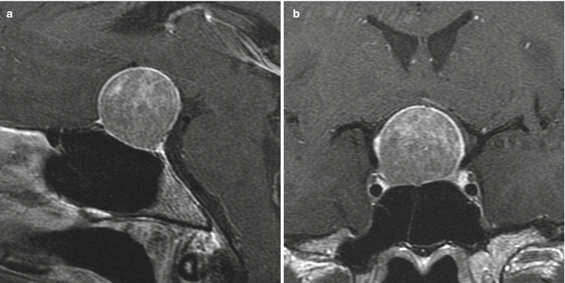
Fig. 10.2
Nonfunctioning pituitary adenoma. (a) Sagittal T1-weighted post-gadolinium image. (b) Coronal T1-weighted post-gadolinium image. There is a heterogeneous, predominantly hypoenhancing sellar mass with remodeling of the sellar floor and suprasellar extension with elevation of the optic chiasm
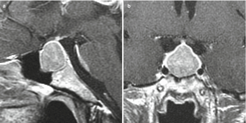
Fig. 10.3
Nonfunctioning pituitary adenoma. (a) Sagittal T1-weighted post-gadolinium image. (b) Coronal T1-weighted post-gadolinium image. A hypoenhancing sellar mass abuts the cavernous internal carotid arteries without encasement. There is suprasellar extension but no significant mass effect on the optic chiasm
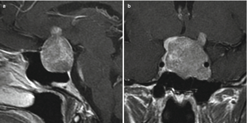
Fig. 10.4
Nonfunctioning pituitary adenoma. (a) Sagittal T1-weighted post-gadolinium image. (b) Coronal T1-weighted post-gadolinium image. There is a hypoenhancing sellar mass with remodeling of the sellar floor. The mass encases the left cavernous internal carotid artery, suggestive of cavernous invasion. The image also shows suprasellar extension with elevation of the optic chiasm, with a small nodular component contacting the left hypothalamus. The normal gland appears along the superior right aspect of the mass
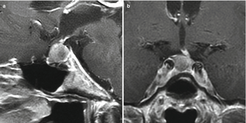
Fig. 10.5
Nonfunctioning pituitary adenoma. (a) Sagittal T1-weighted post-gadolinium image. (b) Coronal T1-weighted post-gadolinium image. There is a hypoenhancing sellar mass abutting the right cavernous internal carotid artery without encasement. The pituitary stalk is deviated to the left
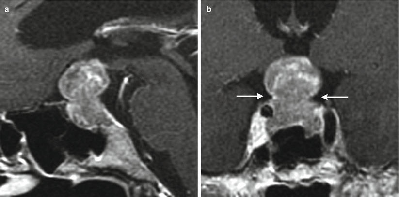
Fig. 10.6
Nonfunctioning pituitary adenoma. (a) Sagittal T1-weighted post-gadolinium image. (b) Coronal T1-weighted post-gadolinium image. A hypoenhancing sellar mass remodels the sellar floor with a nodular component extending to the suprasellar compartment. The mass demonstrates a “snowman” appearance with a waist indicating the level of dural defect or dural thinning (white arrows). The optic chiasm is likely to be superiorly displaced. The mass abuts the cavernous internal carotid arteries without encasement
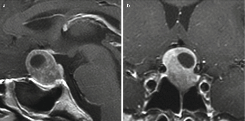
Fig. 10.7
Nonfunctioning pituitary adenoma. (a) Sagittal T1-weighted post-gadolinium image. (b) Coronal T1-weighted post-gadolinium image. There is a hypoenhancing sellar mass with remodeling of the sellar floor and a partly cystic component extending to the suprasellar compartment. The optic chiasm is superiorly displaced
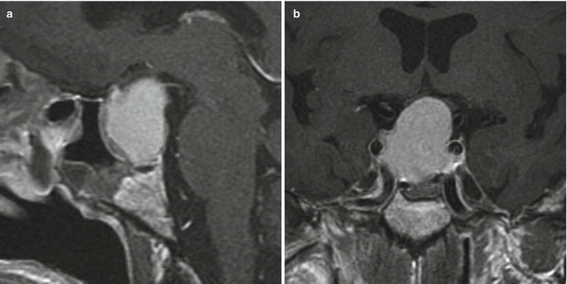
Fig. 10.8
Nonfunctioning pituitary adenoma. (a) Sagittal T1-weighted post-gadolinium image. (b) Coronal T1-weighted post-gadolinium image. A hypoenhancing sellar mass remodels the sellar floor and extends to the suprasellar compartment, displacing the optic chiasm superiorly. The mass abuts the cavernous internal carotid arteries without encasement
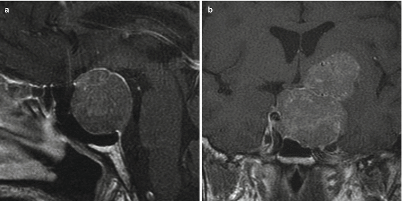
Fig. 10.9
Nonfunctioning pituitary adenoma. (a) Sagittal T1-weighted post-gadolinium image. (b) Coronal T1-weighted post-gadolinium image. There is a hypoenhancing sellar mass with remodeling of the sellar floor and a nodular component extending to the left suprasellar compartment impressing on the left hypothalamus. The mass extends to the left cavernous sinus, lateral to the cavernous internal carotid artery. On the right, the mass abuts the right cavernous internal carotid artery without definite encasement
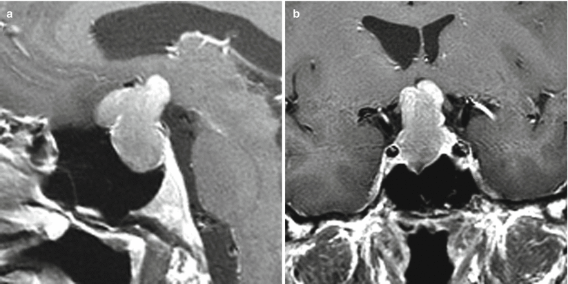
Fig. 10.10
Nonfunctioning pituitary adenoma. (a) Sagittal T1-weighted post-gadolinium image. (b) Coronal T1-weighted post-gadolinium image. A hypoenhancing sellar/suprasellar mass contacts the hypothalamus and displaces optic chiasm superiorly. The mass abuts the right cavernous internal carotid artery without encasement
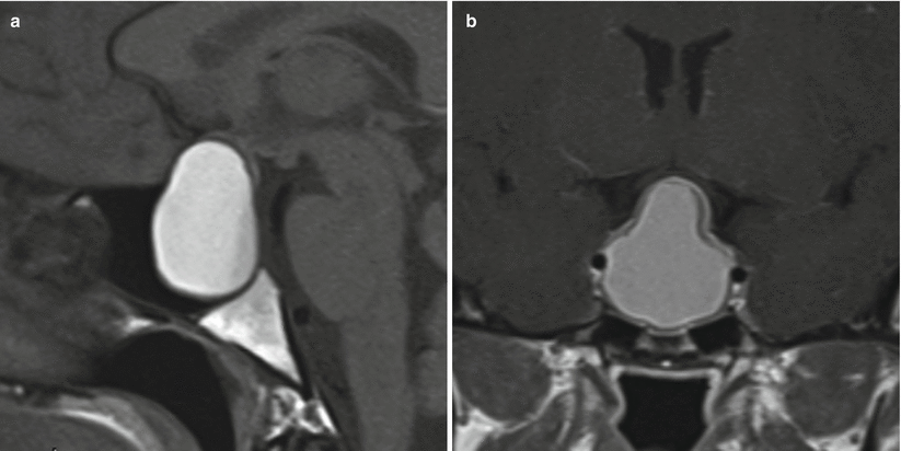
Fig. 10.11
Nonfunctioning pituitary adenoma. (a) Sagittal T1-weighted pre-gadolinium image. (b) Coronal T1-weighted post-gadolinium image. There is a peripherally enhancing sellar mass containing T1 hyperintense content, with remodeling of the sellar floor and a component extending to the suprasellar compartment contacting the hypothalamus. The optic chiasm is superiorly displaced. The mass abuts the cavernous internal carotid arteries without encasement
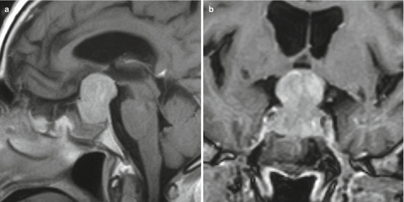
Fig. 10.12
Nonfunctioning pituitary adenoma. (a) Sagittal T1-weighted post-gadolinium image. (b) Coronal T1-weighted post-gadolinium image. A hypoenhancing sellar/suprasellar mass contacts the hypothalamus and displaces the optic chiasm superiorly. The mass abuts the right cavernous internal carotid artery without encasement
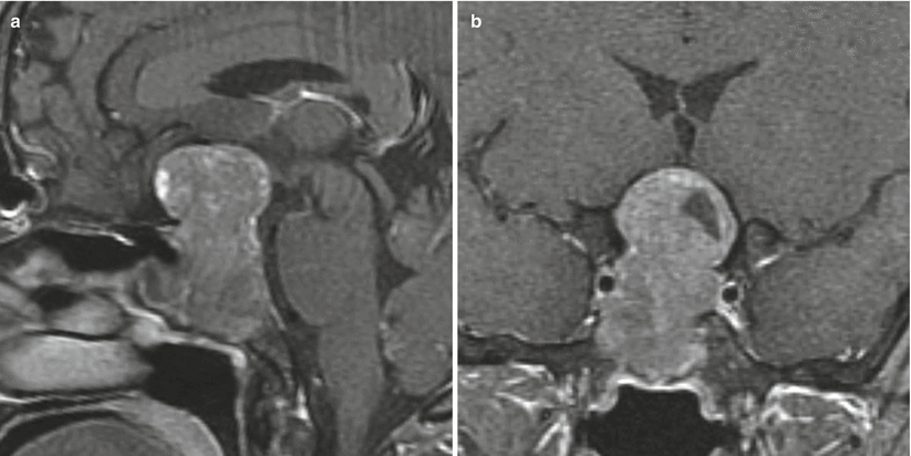
Fig. 10.13
Nonfunctioning pituitary adenoma. (a) Sagittal T1-weighted post-gadolinium image. (b) Coronal T1-weighted post-gadolinium image. There is a heterogeneously enhancing sellar mass with erosion of the sellar floor and a nodular component extending to the suprasellar compartment, contacting the hypothalamus. The optic chiasm is superiorly displaced. The mass abuts the right cavernous internal carotid artery and possibly invades the right cavernous sinus
10.2.2 Histopathology
In 40–50 % of cases, NFPAs demonstrate immunoreactivity for gonadotrope-secreting cells, but it is exceedingly rare for them to secrete functional follicle-stimulating hormone (FSH) or luteinizing hormone (LH) causing clinically apparent changes [4, 7–9]. (See the later section on gonadotroph adenomas.)
Silent FSH/LH adenomas are the most common subtype of surgically resected NFPA. They comprise large cells with a sinusoidal or sheetlike growth pattern that may be interrupted by pseudorosettes and pseudopapillae. Most adenomas stain for both FSH and LH, followed by FSH alone and finally LH alone. Many NFPAs also stain for the alpha subunit [4, 10].
Null-cell adenomas do not stain for any of the anterior pituitary hormone immunostains (Fig. 10.14). They are composed of small to medium cells in sinusoidal or diffuse growth patterns. Chromogranin A is typically strongly expressed and can be used to differentiate them from other intrasellar neoplasms [4, 10].
Tumors staining positive for any hormonal subtype may be “silent.” They comprise 12.6 % of all pituitary adenomas. The most common of these are silent-ACTH (adrenocorticotropic hormone) adenomas, followed by plurihormonal tumors and then silent prolactinomas [4].
Null-cell adenomas are more likely to be atypical adenomas than are silent-gonadotropin adenomas.
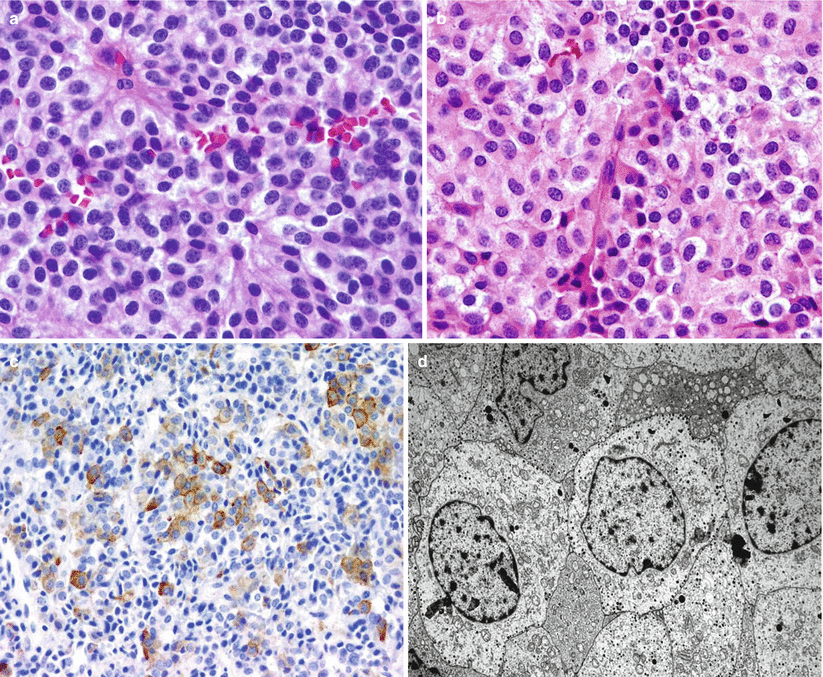
Fig. 10.14


Null-cell adenoma. (a) Null-cell adenomas are usually composed of chromophobic cells arranged in a diffuse or solid growth pattern. (b) Oncocytic changes are not unusual in these adenomas. (c) Although the great majority of null-cell adenomas are immunonegative for any pituitary hormones, focal immunostain for the α-subunit of glycoproteins may be seen. (d) Ultrastructural studies reveal sparse numbers of small, neurosecretory granules with lack of differentiation of any adenohypophyseal specific cell type
Stay updated, free articles. Join our Telegram channel

Full access? Get Clinical Tree


