Other Motor Neuron Diseases and Motor Neuropathies

68 Other Motor Neuron Diseases and Motor Neuropathies


Clinical Vignette


A 63-year-old man was evaluated for 3 years of slowly progressive symptoms of proximal lower extremity weakness. He first noted difficulty negotiating the high step up from the dock to the deck of his boat. Eleven years prior to his neurologic evaluation for weakness, he had been evaluated for painful enlarged breasts, attributed to alcoholic liver disease. More recently, he had noticed difficulty swallowing and had received the Heimlich maneuver on one occasion. Increasingly, muscle cramping had become an annoyance. His mother was troubled by dysphagia and questionable weakness in later life; a brother, two sons, and a daughter had no symptoms. Examination was remarkable for mild facial, tongue, neck flexor and proximal weakness of upper and lower extremities. Tongue and chin fasciculations were seen. There was a mild postural tremor of the hands. Muscle stretch reflexes were traced at the patellar and biceps tendons but otherwise absent. Sensory examination was normal. There were no upper motor neuron signs. Serum creatine kinase level was modestly elevated to 500 IU/L. On nerve conduction studies (NCS), the amplitudes of sensory nerve action potentials were reduced. Needle electromyography (EMG) demonstrated reduced recruitment of high-amplitude and long-duration motor unit potentials consistent with chronic partial denervation and reinnervation in a generalized distribution. Fasciculation potentials were abundant. No evidence of active denervation was demonstrable. Genetic testing revealed expansion of the CAG trinucleotide repeat (>35 CAGs) in the androgen receptor (AR) gene.


Motor neuron diseases (MNDs) are disorders that produce painless weakness, atrophy, cramps, and fasciculations and are consequent to degeneration of anterior horn cells and selective cranial nerve nuclei. This chapter will address notable MNDs other than ALS.


Many of the disorders discussed in this chapter have known or suspected genetic mechanisms. The spinal muscular atrophies (SMAs) are conceptualized as largely inherited disorders in which there is predominant degeneration of anterior horn cells and selective cranial nerve nuclei. In the childhood SMAs, mutations of a single gene and derangement of a single gene product are responsible for the majority of cases, and the resultant phenotype is fairly homogeneous. In other disorders, for example, hereditary spastic paraplegia (HSP), there are a plethora of recognized genotypes correlating with an almost equally heterogeneous array of phenotypic variations.


Because of the relative rarity of these disorders, societal impact is usually limited. However, as with most hereditary disorders, the impact on individuals and families is substantial. This is particularly true for spinal muscular atrophy type I where parents have to cope with the consequences of a newborn with a lethal illness as well as with the specter that subsequent children are at risk. SMA I, also known as Werdnig–Hoffman disease, is the most common of the SMAs. Its incidence is estimated to be between 4 and 10 in 100,000 live births depending on the geographic cohort studied. After cystic fibrosis, it is the second most common, lethal, recessively inherited disorder of Caucasians.


Clinical Presentation


Spinal Muscular Atrophy Types I–IV


Spinal muscular atrophy types I–IV are allelic disorders of the survival motor neuron (SMN) gene 1 located on chromosome 5q12.2-q13.3. When there is more than one affected individual in a given family, the phenotype is typically homogeneous but may be disparate in some cases. In normal individuals, there are two copies each of the SMN1 and SMN2 genes. Both genes produce similar but not identical proteins; the SMN2 gene appears to produce an unstable and rapidly degrading protein that can partially compensate for the lack of the SMN1 protein. There are no known clinical consequences from mutations of the SMN2 gene alone.


It is estimated that 95% of SMA I–III patients are homozygous for deletion of exons 7 and 8 of the SMN1 gene. The remainder are thought to be compound heterozygotes with absence of exons 7 and 8 on one allele and a point mutation of the other SMN1 allele. The severity of the SMA phenotype appears to be related to the number of SMN2 copies available to compensate for deleted SMN1 gene. Homozygotes devoid of SMN1 who harbor two copies of SMN2 tend to manifest as an SMA I phenotype. An increasing number of SMN2 copies correlates with proportionately milder (SMA II-IV) forms of the disease. Individuals homozygous for the SMN1 mutation with five copies of the SMN2 gene have been reported to be asymptomatic. Why motor neurons remain selectively vulnerable to SMN deficiency remains unknown.


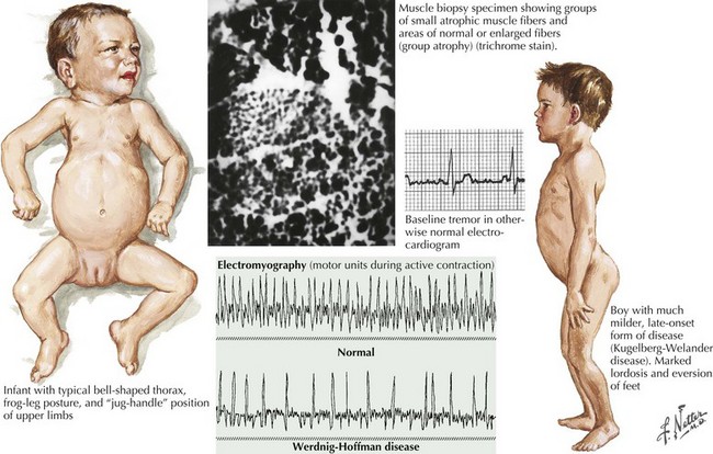
Of the multiple SMA phenotypes, the infantile and childhood forms are the most prevalent. SMA type I or Werdnig–Hoffman disease is the most severe form (Fig. 68-1). Clinical manifestations become evident within the first 6 months of life. In contrast to the latter three categories, afflicted children with SMA I never develop the capability of sitting independently. In some cases, recognition of reduced movement occurs in utero or within the first few days of life. Affected infants are hypotonic with a symmetric, generalized, or proximally predominant pattern of weakness. Like ALS, facial weakness is typically mild and extraocular muscles are spared. Fasciculations are seen in the tongue but rarely in limb muscles, presumably because of the ample subcutaneous tissue of neonates. Manual tremor, so characteristic of SMA types II and III, is rarely present. Deep tendon reflexes are typically absent. Abdominal breathing, a weak cry, and a poor suck are commonplace. Ventilation difficulties stem primarily from intercostal rather than diaphragmatic weakness. Pectus excavatum and a diminished anteroposterior diameter of the chest are seen. Mild contractures may occur but arthrogryposis is not part of the classic phenotype. Intellectual development is normal. Without mechanical ventilation, death is inevitable, almost always within a year or two. An earlier age of onset correlates with a shorter life expectancy.


SMA type Ia refers to a severe form of neonatal SMA associated with arthrogryposis multiplex congenita and a paucity of movement. Prognosis is poor with ventilatory support required at birth.


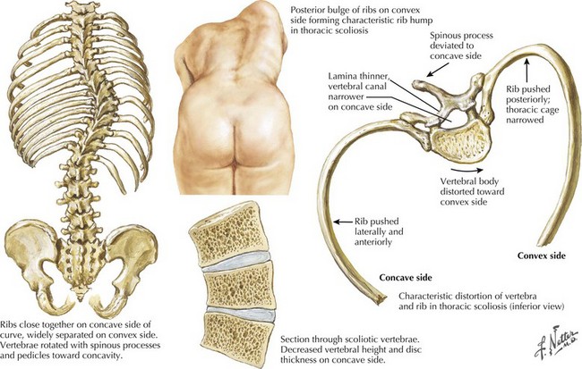
The intermediate form or SMA II typically begins between 6 and 18 months of age. The disorder is clinically defined by a child who sits independently but never walks. Postural hand tremor is the only significant phenotypic variance from Werdnig–Hoffman disease. Tongue fasciculations, areflexia, and a generalized to proximally predominant and symmetric pattern of weakness mimic the SMA I phenotype. Approximately 98% of these individuals survive to age 5 and two thirds to age 25. In view of the more protracted course and of wheelchair dependency SMA II and SMA III patients commonly acquire kyphoscoliosis and joint contractures (Fig. 68-2).


The SMA III or the Kugelberg–Welander syndrome differs from the intermediate form only in the age of onset, milestones achieved, and life expectancy. Affected individuals develop the ability to stand and walk. Onset age is typically 18 months or more. Certain authors have attempted to divide SMA III into type a and type b, based on age at onset of symptoms, with the intention of better defining the natural history in individual patients. In SMA type IIIa, defined as symptom onset before 3 years, it is estimated that 70% will remain ambulatory 10 years after symptom onset. Twenty percent will still ambulate in 30 years after symptom onset. In SMA type IIIb, defined as symptom onset after 3 years, virtually all patients will remain ambulatory in 10 years and 60% at 40 years after symptom onset. Life expectancy extends into the sixth decade and may be normal in many individuals. Initial symptoms are typically related to proximal weakness. Hand tremor, areflexia, and tongue fasciculations are commonplace. Fasciculations in limb muscles are more evident than in SMA types I and II.


Adult-onset SMA IV is a rare, genetically heterogeneous disorder. SMA IV children achieve motor milestones at normal ages. Onset of weakness is typically in the third or fourth decade in the recessively inherited cases. Initial symptoms are typically proximal weakness of the lower extremities, particularly the hip flexors, hip extensors, and knee extensors. Shoulder abductors and elbow extensors are the most frequently affected muscles of the arms. Tongue fasciculations, hand tremor, and in some cases, calf hypertrophy may occur. Life expectancy in SMA IV is normal. Parents with SMA IV have given birth to children with more severe SMA phenotypes.


X-Linked Bulbospinal Muscular Atrophy (SBMA)—Kennedy Disease


SBMA is an X-linked disorder associated with an androgen receptor gene mutation. Consequently, it has frequent endocrine as well as neuromuscular consequences, the latter providing the primary source of morbidity.


SBMA is an X-linked, adult-onset disorder that is depicted in the vignette at the beginning of this chapter. It is a disorder almost exclusively of males with a median age of onset of 44 years. Initial symptoms are usually attributable to weakness of bulbar or proximal limb muscles. Younger men, and rarely female carriers, may be symptomatic but may go undiagnosed unless there are other previously diagnosed family members.


As the name implies, the clinical manifestations are largely referable to degeneration of the lower cranial nerve motor nuclei and anterior horn cells of the spinal cord. The weakness progresses insidiously and is proximally predominant and symmetric in pattern. Typically, symptoms referable to the lower extremities have the greatest initial impact. Approximately 10% of the time, the initial symptoms pertain to difficulty with swallowing, chewing, or speaking. Facial weakness is common. Jaw drop due to muscles of mastication may occur as well. Perioral and tongue fasciculations are common and represent helpful clinical clues. Like ALS, ptosis and ophthalmoparesis should suggest an alternative diagnosis. Like other SMAs, postural tremor is common. There is an associated, but frequently asymptomatic, sensory neuropathy that may only be recognized by nerve conduction studies. Clinical heterogeneity exists. Asymmetry of muscle weakness at onset has been emphasized by some authors. Occasionally, rapidly progressive weakness occurs. The median age of wheelchair dependency is 61 years or approximately 15 years after onset of weakness. Women who are heterozygous for Kennedy disease mutation may rarely be symptomatic.


The effects of SBMA are not restricted to the neuromuscular system. Affected males suffer the consequences of androgen insensitivity, including gynecomastia, impotence, testicular atrophy, and potential infertility. There is also an increased incidence of diabetes mellitus.


Juvenile Segmental Spinal Muscular Atrophy—Benign Focal Amyotrophy—Hirayama Disease


Unlike other SMAs, Hirayama disease appears to be a sporadic disorder in the majority of cases. In 1963, Hirayama described a slowly progressive, focal motor neuron disease affecting one, and at times, both upper extremities. In this and subsequent descriptions, males are affected in 60% of cases. Hirayama disease is perhaps best considered as a segmental or regional form of spinal muscular atrophy. Onset is typically between ages 15 and 25 with a range of 2 to 30 years. Although most commonly reported in those of Asian origin, it may occur in any ethnic background.


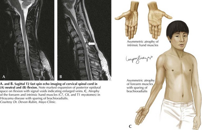
The characteristic phenotype is the insidious development of atrophy and weakness in C8–T1 muscles of the hand and forearm. It begins unilaterally, typically in the dominant extremity. Over the course of months to years, the weakness may gradually spread to involve more proximal muscles. In a third of cases, there is clinical weakness of the opposite limb. An even higher percentage will have bilateral upper extremity involvement on electrodiagnostic studies. Tendon reflexes in the involved limb may be spared, although neither overt pyramidal or bulbar involvement occurs. Reflex preservation may reflect the restricted nature of the disease and the lack of a reliable C8–T1 muscle stretch reflex. Like many other SMAs, tremor may occur. In most cases, there is an arrest of further progression after 6 years or less. Although a significant decline in affected limb function in the cold is common with all motor neuron diseases, “cold paresis” is particularly emphasized in this population. Hyperhidrosis of the involved limb has been described.


Hirayama disease is less frequently seen in Western populations. Ischemic changes in the cervical spinal cord of a single autopsied case of Hirayama disease led to the hypothesis of a compressive mechanism. In 2000, Hirayama reported the results of dynamic imaging in 73 patients and 20 controls. Ninety-four percent of patients had significant forward displacement and flattening of the posterior surface of the cervical cord during neck flexion (Fig. 68-3). The presumption is that the blood supply to the spinal cord is compromised, with the anterior horn representing the watershed and the most susceptible to ischemia. Other observations that supported this potential mechanism are the frequent asymmetric nature of spinal cord flattening in keeping with the asymmetric disease onset, and the lesser degree to which distortion occurred in older patients in whom progression had arrested. Nonetheless, this pathogenetic hypothesis is not universally accepted.
Jun 4, 2016 | Posted by in NEUROLOGY | Comments Off on Other Motor Neuron Diseases and Motor Neuropathies

Full access? Get Clinical Tree

Get Clinical Tree app for offline access