Fig. 22.1
Rathke cleft cyst. (a) Sagittal T1-weighted post-gadolinium image. (b) Coronal T1-weighted post-gadolinium image. A homogeneous, nonenhancing lesion is seen in the posterior sella, displacing the pituitary gland anteriorly. The pituitary stalk mildly deviates to the left
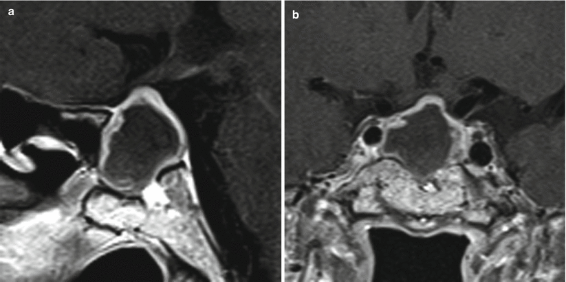
Fig. 22.2
Rathke cleft cyst. (a) Sagittal T1-weighted post-gadolinium image. (b) Coronal T1-weighted post-gadolinium image. There is a cystic lesion with peripheral enhancement in the sella, with erosion of the sellar floor and extension to the suprasellar cistern, displacing the optic chiasm
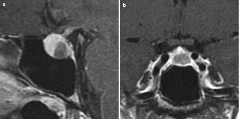
Fig. 22.3
Rathke cleft cyst. (a) Sagittal T1-weighted post-gadolinium image. (b) Coronal T1-weighted post-gadolinium image. A cystic lesion is centered in the posterior sella, displacing the pituitary gland anteriorly and superiorly
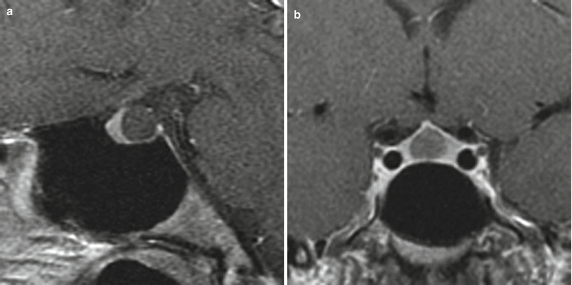
Fig. 22.4
Rathke cleft cyst. (a) Sagittal T1-weighted post-gadolinium image. (b) Coronal T1-weighted post-gadolinium image. A cystic lesion is located in the posterior sella, displacing the pituitary gland anteriorly
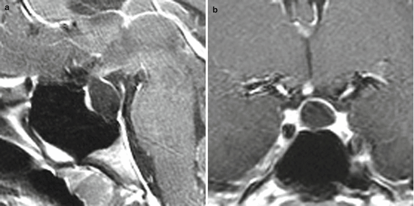
Fig. 22.5
Rathke cleft cyst. (a) Sagittal T1-weighted post-gadolinium image. (b) Coronal T1-weighted post-gadolinium image. There is a cystic lesion in the posterior sella, displacing the pituitary gland anteriorly. There is remodeling of the sellar floor
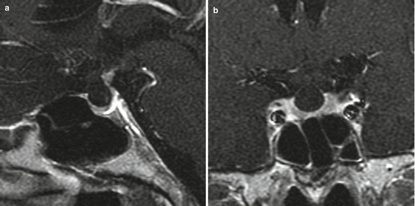
Fig. 22.6
Rathke cleft cyst. (a) Sagittal T1-weighted post-gadolinium image. (b) Coronal T1-weighted post-gadolinium image. A cystic sellar lesion displaces the pituitary gland inferiorly and the stalk posteriorly
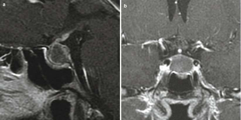
Fig. 22.7
Rathke cleft cyst. (a) Sagittal T1-weighted post-gadolinium image. (b) Coronal T1-weighted post-gadolinium image. There is a cystic lesion in the posterior sella displacing the pituitary gland and stalk anteriorly. There is remodeling of the sellar floor
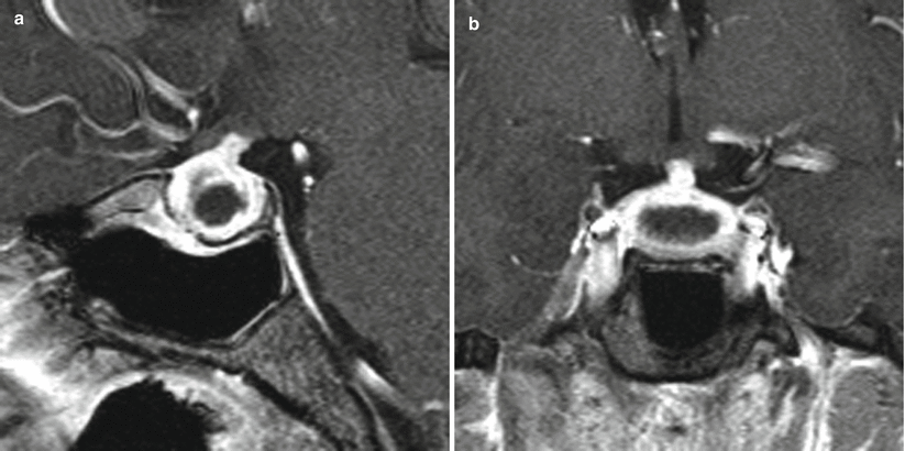
Fig. 22.8
Rathke cleft cyst. (a) Sagittal T1-weighted post-gadolinium image. (b) Coronal T1-weighted post-gadolinium image. A cystic lesion within the sella displaces the stalk anteriorly. There is remodeling of the sellar floor
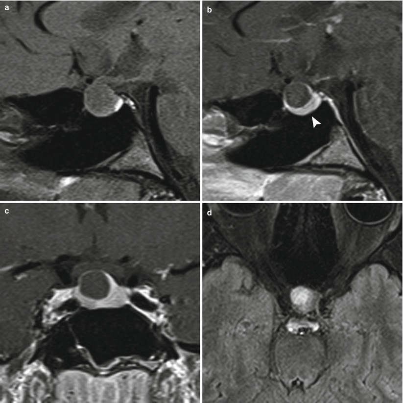
Fig. 22.9
Rathke cleft cyst. (a) Sagittal T1-weighted pre-gadolinium image. (b) Sagittal T1-weighted post-gadolinium image white arrow points to normal pituitary. (c) Coronal T1-weighted post-gadolinium image. (d) Axial FLuid Attenuated Inversion Recovery (FLAIR) image. There is a cystic lesion in the sella, displacing the pituitary stalk anteriorly. There is remodeling of the sellar floor. The optic chiasm is mildly elevated
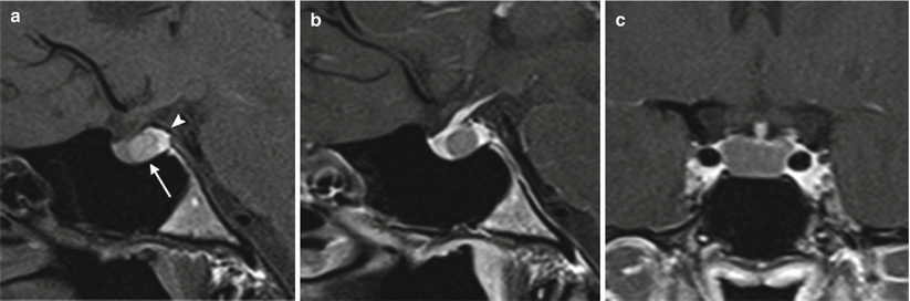
Fig. 22.10
Rathke cleft cyst. (a) Sagittal T1-weighted pre-gadolinium image. (b) Sagittal T1-weighted post-gadolinium image. (c) Coronal T1-weighted post-gadolinium image. A homogeneous, cystic lesion (arrow) contains T1 hyperintense material between the adenohypophysis and neurohypophysis (arrowhead)
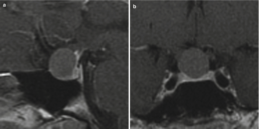
Fig. 22.11
Rathke cleft cyst. (a) Sagittal T1-weighted post-gadolinium image. (b) Coronal T1-weighted post-gadolinium image. There is a cystic lesion in the anterior sella, displacing the pituitary gland inferiorly and the pituitary stalk posteriorly
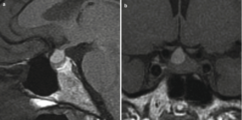
Fig. 22.12
Rathke cleft cyst. (a) Sagittal T1-weighted pre-gadolinium image. (b) Coronal T1-weighted pre-gadolinium image. A cystic lesion contains T1 hyperintense material in the anterior sella, displacing the pituitary gland inferiorly
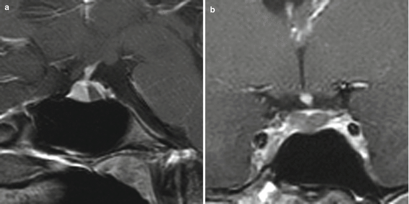
Fig. 22.13
Rathke cleft cyst. (a) Sagittal T1-weighted post-gadolinium image. (b) Coronal T1-weighted post-gadolinium image. A homogeneous, nonenhancing lesion is located between the adenohypophysis and neurohypophysis
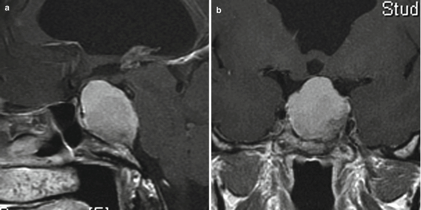
Fig. 22.14
Rathke cleft cyst. (a) Sagittal T1-weighted post-gadolinium image. (b) Coronal T1-weighted post-gadolinium image. There is a large, T1 hyperintense lesion eroding the sellar floor and extending to the suprasellar cistern, mildly elevating the optic chiasm. The mass extends posteriorly, abutting the pons
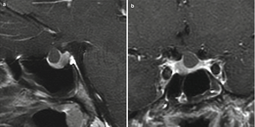
Fig. 22.15
Rathke cleft cyst. (a) Sagittal T1-weighted post-gadolinium image. (b) Coronal T1-weighted post-gadolinium image. There is a cystic lesion in the anterior sella, displacing the pituitary gland inferiorly. In addition, there is a hypoenhancing mass in the right aspect of the sella, likely to represent an incidental pituitary adenoma
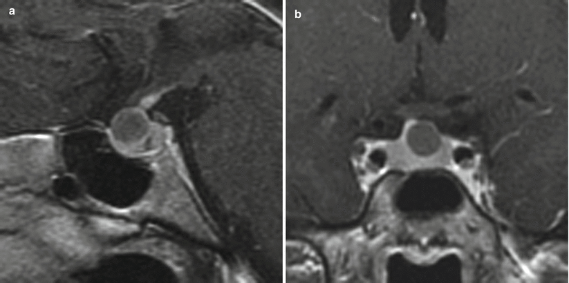
Fig. 22.16
Rathke cleft cyst. (a) Sagittal T1-weighted post-gadolinium image. (b) Coronal T1-weighted post-gadolinium image. A cystic lesion is present in the anterior sella, displacing the pituitary gland inferiorly. The pituitary stalk is deviated to the right
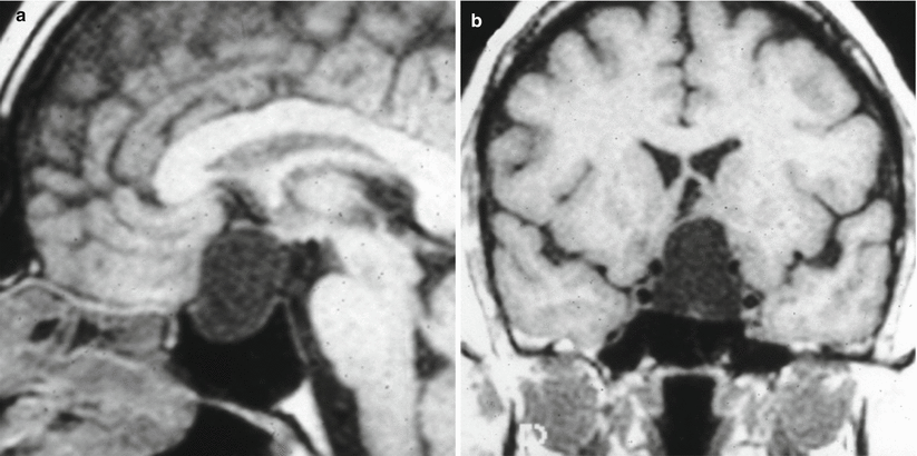
Fig. 22.17
Rathke cleft cyst. (a) Sagittal T1-weighted pre-gadolinium image. (b) Coronal T1-weighted pre-gadolinium image. A cystic sellar/suprasellar lesion elevates the left hypothalamus
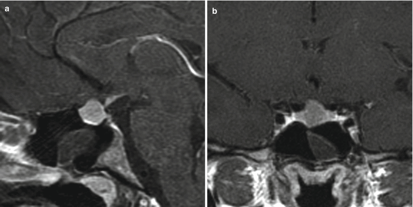
Fig. 22.18
Rathke cleft cyst. (a) Sagittal T1-weighted pre-gadolinium image. (b) Coronal T1-weighted pre-gadolinium image. There is a nonenhancing, T1 hyperintense sellar lesion with mild suprasellar extension
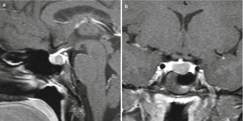
Fig. 22.19
Rathke cleft cyst. (a) Sagittal T1-weighted post-gadolinium image. (b) Coronal T1-weighted post-gadolinium image white arrow. A cystic lesion is centered between the adenohypophysis and neurohypophysis. The lesion is posterior and inferior to the pituitary stalk
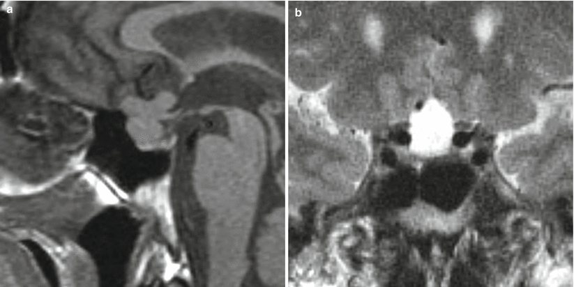
Fig. 22.20


Rathke cleft cyst. (a) Sagittal T1-weighted pre-gadolinium image. (b) Coronal T2-weighted image. There is a cystic lesion in the sella with superior extension to the anterior interhemispheric fissure
Related posts:


Stay updated, free articles. Join our Telegram channel
Full access? Get Clinical Tree
