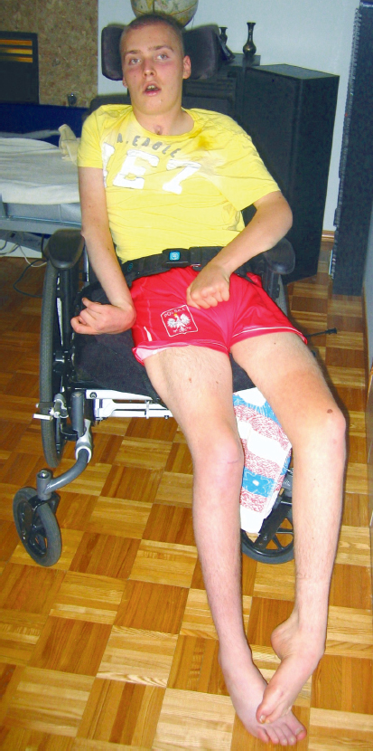
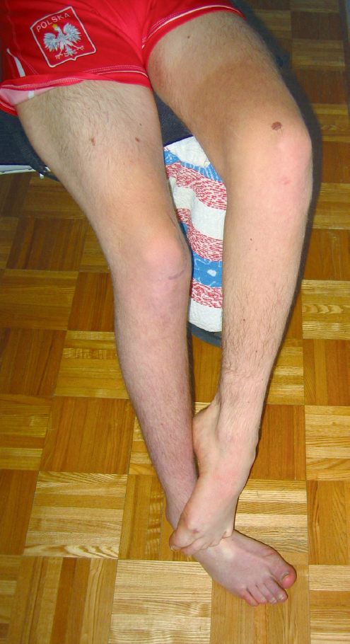
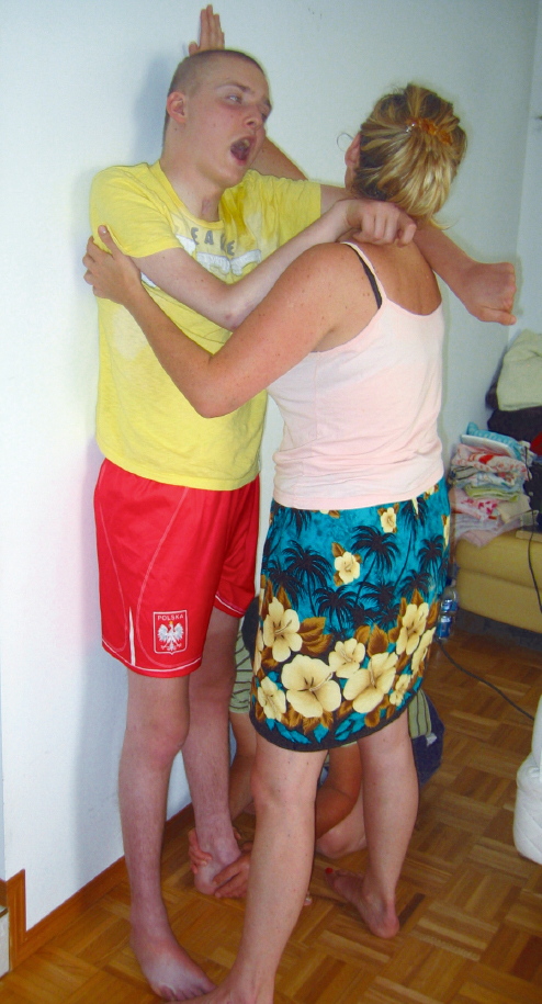
Case Report A4 Addressing the Primary and Secondary Impairments of a 20-Year-Old Man with Traumatic Brain Injury Severe traumatic brain injury (TBI) may cause primary impairments, including reduced (or absent) muscle activation and motor execution as well as spasticity.1 Depending on the severity and the conditions of the acute and subacute recovery phases, years of immobility and malalignments may be incurred and can lead to significant secondary impairments. Complete and partial bed rest, as a medical order and as a by-product of resources required to mobilize, is often an issue in the acute and subacute phases postinjury. However, immobilization of body segments, such as the affected feet and ankles or wrists and hands, is an ongoing concern. Unfortunately, well-meaning therapists often contribute to this immobilization with splints, braces, and positioning devices.2,3 Secondary consequences of the primary impairments and ensuing immobilization include joint deformities, muscle atrophy, soft tissue composition changes, further sensory changes, and cardiorespiratory compromise.4 Physical therapists (PTs) and occupational therapists (OTs) understand these effects of immobility on the various systems of the human body, yet practitioners often intervene with these systems in isolation, focusing on one issue at a time. This case report reviews the recovery of an individual with a severe TBI, highlighting the significant relationship between the musculoskeletal system and the neurological system. The muscles and joints of the ankles and feet are commonly affected by spasticity, overrecruitment or overflow activity, poor positioning, and immobility. Maintaining normal alignment and mobility in these areas is crucial for shock absorption, balance reactions, efficient weight bearing, and sensory feedback.5 In addition, the foot and ankle complex influence the biomechanics of other joints and muscles up the kinetic chain because adults spend the majority of their day functioning in upright positions (sitting, standing, and walking activities). Neuro-Developmental Treatment (NDT) principles can be used in conjunction with orthopedic knowledge and strategies to address not only the primary impairments caused by TBI but the secondary joint and soft tissue issues related to reduced, absent, or abnormal movement patterns. Ernie is a 20-year-old man who suffered a TBI as a result of a motorbike accident (MBA). He was 17 years of age at the time of his accident and was a healthy, active high school student who enjoyed history, politics, and snow-boarding. The MBA left him with bifrontal contusions, a diffuse axonal brain injury, and a large arachnoid cyst. In addition, he had a left wrist fracture and multiple fractures of the spine with no spinal cord involvement. His family was given a very poor prognosis for any recovery of cognitive or physical function. Although Ernie’s family understood the prognosis, they chose to be supportive, hardworking, and devoted to any recovery he could achieve. As such, 15 months after the MBA, the family contacted an NDT-certified PT. This case study began in Ernie’s home, after he had already received intervention in an acute neurology unit, rehabilitation from an inpatient unit, and a brief experience with an out-patient program. Intervention took place in his home once a week for 8 months, then once a month for ~ 5 months, and then once every few months until a total of 21 months had passed from our first meeting. This home-based functional physiotherapy approach, combined with the dedication of Ernie’s family and home care worker in following through with recommended activities, ensured regular practice and thus his recovery and success. The expressed goals of Ernie and his family were to decrease the amount of assistance required for transfers and mobility, as well as to increase his tolerance for activities and improve his quality of life (i.e., standing and walking activities). Ernie lives at home with his mother, father, and older brother. The family converted the television room on the main floor of the home into a bedroom for him. He had not been up the 12 stairs to his previous bedroom since before his TBI. Because the television room is sunken, the family built a ramp for Ernie’s access. This ramp can be moved to the step into the kitchen or to the step up to the bathroom. Ernie is completely dependent for all of his activities of daily living (dressing, bathing, toileting, feeding) and mobility. He has been wheelchair dependent since the accident (Fig. A4.1). Ernie’s mother first positioned him in sitting, close to the edge of the bed. Ernie attempted to help with some of the movements, mostly using a forward head and neck pattern and pulling his left upper extremity (UE) toward adduction and internal rotation. In sitting, he remained in a posterior pelvic tilt with a significantly rounded low back and thoracic spine. As a result, his neck was in suboccipital extension with his head dropping to the left and down to his chest. Fig. A4.1 Ernie in a common wheelchair sitting position early in his intervention program. Ernie’s family and caregivers attempted to place his feet under his knees in preparation for the transfer. However, due to an equinovarus position, he was unable to bear weight on the plantar surfaces of his feet (Fig. A4.2). His weight-bearing surfaces were primarily the lateral borders of his fourth and fifth metatarsals toward their metatarsal heads. Left ankle dorsiflexion (after realignment and overpressure) was approximately − 8° and right was approximately − 10°. One person was in front of Ernie to move him forward over his feet and help with lifting his buttocks off the bed. The second person to assist with the transfer was behind Ernie, helping to lift and guide his hips to the chair/wheelchair. The transfer was performed quickly, and Ernie remained passive during the movement. Throughout the transfer, his entire trunk was in flexion, including the cervical, thoracic, and lumbar areas. His ankles never achieved neutral alignment (in either dorsiflexion/plan-tar flexion or inversion/eversion). Fig. A4.2 The equinovarus position of Ernie’s bilateral lower extremities. Ernie stood with three people supporting him. One person was at Ernie’s right lower extremity (LE) to ensure that his knee neither hyperextended nor buckled into flexion. A second person was required at the left leg to limit the overactivity that pushed him into extension and to the right. Repositioning of the feet was required to ensure at least partial weight bearing on the plantar aspects of his feet and to prevent injury to Ernie’s ankles. As such, his feet tended to be placed far apart (hip abduction) and in toe-out positions (hip external rotation), increasing the width of his base of support (BOS) to ~ 35 cm (13.75 in) measured from the third digit of each foot. The third person was behind Ernie, in high kneeling on the bed, and supported him through his chest and shoulders, while also providing assistance at his gluteal muscles and hips to maintain hip extension. Later, a wall was used to support Ernie’s trunk in standing, and the third person could then come in front of him and talk with him to work on head and neck alignment and control. Ernie’s alignment was with trunk and hips behind his BOS and to the right (Fig. A4.3). Table A4.1 outlines Ernie’s level of functioning within the International Classification of Functioning, Disability and Health (ICF) framework at the time of initial evaluation. It is worth noting the additional positive features that contributed to his ongoing practice and success over time: • Motivation. • Body awareness. • Strong family and friend support systems. • Knowledgeable and dedicated home care support worker. • Overlengthened trunk extensors. • Weak trunk muscles; extensors > flexors. • Unable to sustain activity in trunk extensors and flexors for stable, coactivated trunk. • Tight but not fixed interspinal segments. • Weak cervical muscles; extensors > flexors. • Inability to sustain activity in cervical extensors; long > short for stable, coactivated cervical spine. • Shortened neck side flexors on R (scalene muscles and sternocleidomastoid). Fig. A4.3 Ernie’s standing against a wall required three people for safety and alignment (photographer assisted at right foot and ankle). Note that Ernie’s feet are toed out, abducted, and 18 cm (7 in) from the wall to accommodate the equinovarus restrictions. • Weakness of oral-motor muscles. • Inability to sustain activity in oral-motor muscles. • Recruiting most muscles in a cocontracting manner (agonist and antagonist contracting simultaneously) • Inability to separate and grade muscle activity between agonist and antagonist, as well as synergistically. • Weakness and inability to sustain activity in all left UE muscle groups. • Tightness in long finger flexors. Table A4.1 Using the International Classification of Functioning, Disability and Health (ICF) to outline initial clinical evaluation
A4.1 Introduction
A4.2 The Clinical Relationship between the Musculoskeletal System and the Nervous System
A4.3 Case Description
A4.3.1 Examination
Client and Family Goals
Baseline Functional Performance
Wheelchair to Bed Transfer
Standing for Dressing/Fixing Pants
Summary of Most Significant Single-System Impairments of the Neurological and Musculoskeletal Systems
Trunk
Head
Oral motor
Left UE
Participation/restrictions | Functional activity/limitations | Observations of posture and movement |
• Dependent for all mobility in home and community, including ambulation and transfers. Mother pushed manual wheelchair. | • Unable to sit without one-person assist on typical surfaces. | • Sat or stood in trunk flexion with post–pelvic tilt and suboccipital extension. |
• Required full wheelchair back support. | • Complained of pain and fatigue. | |
• Required full assist of two people for low-pivot transfers. | • Hyperextended left knee in standing. | |
• Required full assist of three people for standing (one on either side, one behind). | • Pushed into extension and to weaker right LE by overactivating left leg muscles. | |
• Left pelvis in posterior rotation; hips behind BOS. | ||
• Left foot and ankle pulled into equinovarus pattern of ankle when standing (plantar flexion, inversion, talus abduction). | ||
• In sitting, left calcaneus rested in inversion, talus in abduction, with ankle in plantar flexion. | ||
• Able to actively move partially out of equinovarus posture for better positioning in wheelchair or in preparation for transfer. | ||
• Left passive dorsiflexion to neutral, with knee flexed and significant overpressure. | ||
• Unable to engage in eye contact with family, friends, or therapists during conversations. | • Inability to right head on body. | • When one person supported head, neck pulled into position of suboccipital hyperextension. |
• Required one person assist to hold head up. | ||
• Unable to scan his environment. | • Head fell to left and down to chest when not supported. | |
• Unable to eat out with family and friends. | • Unable to manage oral secretions. | • Mouth remained open with jaw dropping down; became wider with excitement or frustration. |
• Frustrated by family and friends using cloths to wipe away drool. | • Difficulty closing jaw. | |
• Required one person assist for managing oral care. | • Drooled. | |
• Unable to bite or chew food independently. | ||
• Required one person assist for providing appropriate texture to diet, as well as feeding Ernie. | ||
• Unable to verbally communicate wants or needs—increased time and effort to communicate. | • Unable to show many facial expressions. | • Unable to gesture with right UE. |
• Required one person to ask specific, single-sentence questions. | • Able to reach with left hand for a “high five.” | |
• Communicated with left thumb extension for “yes,”, pinky extension for “no.” | ||
• Gestured “hello” by raising left arm up (to head height). | ||
• Dependent for all basic activities of daily living. | • Unable to use left UE in functional reach (e.g., food, drink, or grooming). | • Able to partially relax tone in right UE to command for positioning and comfort. |
• Unable to grasp or manipulate objects (i.e., open lids on jars, operate pens, hold pictures). | • Rested in a position of significant humeral IR with forearm pronation, wrist and finger flexion. | |
• Unable to use right UE for function. | • Elbow often extended. | |
• Unable to use right UE for bilateral activities. | ||
• Unable to participate when ambulatory activities required. | • Unable to safely use (i.e., at risk of ankle or knee injury) right LE for bilateral weight-bearing activities, such as standing transfers, standing activities, or walking. | • When standing, locks right knee, or knee gives way into flexion. |
• Minimal active weight bearing through right LE. | ||
• Hips behind BOS in standing. | ||
• Caregivers concerned he may sprain his ankle during transfers or standing activities. | • Right ankle resting in equinovarus posture for prolonged periods of time. | |
• Unable to actively recruit right dorsiflexors for positioning or preparation for function. | ||
• Passive right dorsiflexion to − 10° with knee flexed and significant overpressure. |
Abbreviations: BOS, base of support; IR, internal rotation; LE, lower extremity; UE, upper extremity.
Right UE
• Weakness of bicep and pectoralis major muscles—only volitional flickers observed.
• Shortened long finger flexors.
• Inability to initiate activity in the other right UE muscles.
Left LE
• Shortened gastrocnemius/soleus complex, as well as other soft tissues surrounding foot and ankle joints.
• Weakness in left knee flexors, hip extensors and abductors, and ankle evertors and dorsiflexors.
• Inability to selectively recruit and grade muscles within the left leg (between flexors and extensors, between abductors and adductors).
Right LE
• Shortened gastrocnemius/soleus complex, as well as other soft tissues surrounding all foot and ankle joints.
• Weakness of all right LE extensors and flexors.
• Inability to sustain activity of all LE muscles.
A4.3.2 Evaluation and Plan of Care
Limited time for intervention (1 hour a week with an NDT-trained therapist), environment (his home), and stage of recovery (15 months post-TBI) demanded that Ernie’s care plan be prioritized and function-based. There were expectations that the family and home care worker would be able to follow through with activities, as well as ensuring that Ernie remained motivated and engaged in the activities. To achieve both of these criteria, NDT principles of active, upright, and loaded (weight-bearing) positions while incorporating the more affected extremities during meaningful tasks were used.
To achieve these upright, weight-bearing positions, however, required better alignment of the LEs. Normal movement cannot occur in the presence of abnormal alignment.6 As such, priority was placed on stretching, mobilizing, and realigning Ernie’s feet and ankles using both orthopedic manual therapy strategies and functional postures and activities that used his body weight to assist with LE stretching while he was engaged in functional activities.
The significant malalignments of Ernie’s feet and ankles were hypothesized to be primarily due to changes to the length of muscles and soft tissues. There were certainly adhesions of his joints and tendons; however, there were no fixed contractures or areas of heterotopic ossification (hard or bony end-feels). Overrecruitment of his plantar flexors and invertors caused his ankles and feet to assume the equinovarus position for the majority of the day. This prolonged poor positioning caused shortening of the intrinsic flexor foot muscles, plantar flexors (gastrocnemius–soleus complex) and invertors (tibialis posterior), and overlengthening of the dorsiflexors (tibialis anterior) and evertors (peroneals), thus making recruitment of the latter two muscle groups difficult.
The equinovarus positioning contributed to the poor alignment and impairments in muscles throughout the entire lower extremities, trunk, and upper extremities. Ernie stood with his left knee in hyperextension and his left pelvis/hip in posterior rotation. He had contraversive pushing tendencies and pushed into the lateral border and toes of his left foot. Thus his trunk was shifted to the right and back. However, the right leg was unable to support the weight, and he would either hyperextend the right knee for stability or it would collapse into flexion due to weakness. When in contact with the ground, the weight-bearing surfaces of the right foot were primarily the toes (third through fifth digits) and the lateral border of the foot.
As a result of this unstable BOS, asymmetrical alignment, and unequal weight bearing, Ernie did not feel safe or balanced. This led to even more overactivation of his left LE extensors and plantar flexors, feeding into the asymmetrical alignments, and causing the family and caregivers to provide significant support to him. Everyone, especially Ernie, was frustrated and fatigued.
A4.3.3 Intervention
Although the focus was on realigning the feet and ankles, the other impairments were not forgotten. The NDT Practice Model advocates addressing biomechanical and kinesiological principles to achieve the best possible alignment and conditions for appropriate muscle activity. Intervention began with closed chain activities and functional and meaningful movement patterns to achieve increased sensory feedback through joint compression and mechanoreceptor stimulation. It was important to activate the muscles of Ernie’s LEs within normal synergies while performing the stretches and mobilizations to his feet and ankles. Active, closed chain positions balance agonist/antagonist activity, load the individual’s own body weight into the limb, and use eccentric/concentric contractions.7 In addition, the vertical orientation stimulates the reticular activating system, which increases the level of alertness and was motivating to Ernie.
Table A4.2 outlines the activities chosen and the progressions made for achieving the therapy priority of foot and ankle realignment during active, upright, and weight-bearing positions to allow Ernie to transfer more safely and with less assistance. Most tasks were performed with bare feet to allow the family and the therapist direct contact with his ankles and feet for safe alignments and facilitation (including mobilization of the joints and soft tissues) (Fig. A4.4, Fig. A4.5). All movements and activities were attempted from a position of talar neutral.8 The feet were then placed on the floor while maintaining this alignment. Some ankle plantar flexion (PF) was necessary. However, due to hypertonicity and/or short plantar flexors.
Table A4.2 Activities performed during therapy including alignments and handling
Position | Activities performed | Handling/assistance provided |
Sitting on edge of bed with UEs resting on the back of a wingback chair placed in front of Ernie. Placing his UEs on a surface increased his trunk activity and stability. To ensure a stable sitting surface, a 1-in piece of solid wood was placed on top of the mattress for Ernie to sit on. • Bare feet. | Looking at pictures of himself, his family, and friends. The pictures were positioned to encourage weight shift forward to the point of liftoff (lift pelvis/hips offbed to load feet and ankles). • Hold as long as tolerated (goal = 15 s). • Varied the amount of knee flexion (how high lifted up), the duration of the hold and the speed of liftoffand lowering. This variety ensured the type of muscle contraction was adjusted (concentric, eccentric, or isometric) and the ROM demands on the ankle and foot were altered. | • One person to facilitate forward weight shift over UEs and liftoff. • One person at each of right and left feet to ensure increased dorsiflexion as body weight shifted over foot (keep heels on floor) while providing calcaneal and navicular pronation mobilization/hold. |
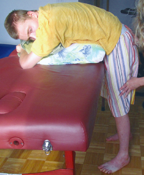
Prone standing (see Fig. A4.4)—using a massage table at highest setting, two pillows under Ernie’s chest as he rests torso and UEs on massage table. Prone standing allows for loading and challenging of the LEs while reducing the demands on the trunk and LEs. Thus concerns related to balance or safety and the amount of work required of helpers are also reduced. • Bare feet. | • Standing with knees slightly flexed for as long as tolerated (for prolonged stretch to ankle plantar flexors [specifically soleus], invertors, and other soft tissues). • Progressively disadvantaged his less involved left LE, first in modified closed chain setups and then to open chain activities. Examples include tapped left heel (unable to toe tap), wiggled left foot out/in, step left foot out/in, step left foot forward/backward. This decreased the amount of pushing activity from the left LE, while increasing the demand and active loading activity of the right, weaker LE. | • One person required at Ernie’s head and torso to ensure comfort and safety (neck position, breathing, swallowing, and UE position) • One person at each of right and left feet to ensure maintenance of ankle dorsiflexion (keep heels on floor) while:
|
Standing against wall introduced at 18 weeks. • Heels placed ~1 in away from wall, BOS from third digit of each foot = 35 cm (13.75 in), knees flexed ~20°. • Bare feet. | Looking at pictures of himself, his family, friends, and motorbike (before and after the accident). Looking at brother, father, or mother when spoken to. • Weight shifting between right and left lower extremities—allowing Ernie to feel off balance to right with left LE pushing, then finding midline and recognizing this midline alignment and balance point. • Wall slides/squats to 20–45° knee flexion, hold for varying lengths of time; again, this modification encourages different types of muscle contractions through various ranges of motion. • Promote loading of more involved right LE during left heel tapping, wiggling left foot out/in. This also helped to decrease the amount of overactivity from left leg extensors (as was done in prone standing). | • Provided extrinsic verbal feedback to improve his awareness of being off midline as well as to improve his correction back to midline (i.e., he often overcorrected alignment and leaned to left with trunk but kept pelvis to right). • Downward and slightly medial physical cue to right gluteus medius muscle to increase, and then sustain, activity in hip abductors (to prevent collapsing into right Trendelenburg). • One person in front for trunk and head alignment and to provide physical cues to left LE (proximal cue at pelvis to maintain midline alignment and to quadriceps to load down into the foot) to decrease hip and knee extensor activity and ankle/foot plantar flexion pushing. • One person at each of right and left feet to ensure maintenance of ankle dorsiflexion (keep heels on floor) while providing calca-neal and navicular pronation mobilization/hold (see Fig. A4.5). |
Standing, no wall support. He was able to begin standing without the wall for support after ~22 weeks. • Bare feet | Looking at pictures of family, friends, and his motorcycle to encourage righting head on body. Moved pictures to various positions of right, center, and left to further challenge balance and trunk control. • Shifted pelvis then shoulders and head forward offwall for brief periods. • Hold with verbal plumbline alignment as tolerated (15–30 s). | • One person required for torso support (through scapula and thoracic spine) as well as to assist with lifting head (verbal and physical cues to look forward at family member—father or brother, or at pictures). • One person at each of right and left feet to ensure maintenance of ankle dorsiflexion (keep heels on floor) while providing calca-neal and navicular pronation mobilization/hold (see Fig. A4.5). |
• Socks, left CaligaLoc (ankle brace), and shoes were worn for first 6–7 months. • Bare feet or shoes (no brace) after ~ 7 months to the end of this case report intervention. | • Assisted weight shift forward to right LE midstance to terminal stance, step with left. Weight shift forward to left LE midstance to terminal stance, step with right. Repeat for ~15 ft, from front door of home to kitchen, over hardwood floor. | • One person in front of Ernie for trunk support and at posterior hips to facilitate forward weight shift to attain, and then maintain, midstance alignment at hips. • One person on each LE to ensure stable foot and ankle alignment during stance phase of gait; reasonable step length during swing; as aligned as possible initial contact to foot flat position for loading; support at knee as required (right knee required more than left to ensure knee flexion without buckling during loading, and soft extension in midstance). • Fourth person to follow with wheelchair for safety. |
Stepups Introduced after ~ 8 months • Bare feet | Using the 6 in step from the sunken living area to the bathroom. • Weight shift forward to attain midstance on right LE, step up with left leg (began with disadvantaging left and increasing active loading response on right LE). • Maintain midstance on right LE without transferring weight onto left LE and step left foot back to floor again (continue to disadvantage the overactive left leg). Repeat as tolerated (initially 5 reps). • Weight shift forward to achieve left midstance, step up with right LE. • Maintain midstance on left LE without transferring weight onto right LE and step right foot back to floor again. Repeat as tolerated (initially 10 reps). | • One person in front of Ernie for trunk support and to facilitate slight forward weight shift to midstance alignment, then assist with maintenance of this position. • UEs around front assister’s waist or shoulders. • One person on each LE to ensure stable foot and ankle of supporting limb; facilitation at stance knee as required (right knee required more than left); appropriate hip and knee flexion during stepups. Ernie was able to initiate left swing, but required moderate assistance to prevent hip hiking and ensure enough knee flexion to clear his toe from the step. Ernie could initiate right swing by releasing the knee, but due to severe weakness, required maximum assistance to lift the weight of his right leg to step up. |
Ascending and descending stairs performed after 9 months of therapy. • Socks and shoes worn. • Left CaligaLoc (ankle brace) worn. | Ascending • Weight shift forward to right midstance alignment, step up to first step (6 in) with left. • Transfer weight forward onto left LE, lift right foot up with heel lifting first, then flex knee and hip, and dorsiflex ankle as lift right foot up to same step. • Repeated above sequence for ascending 12 steps—that is, used a step-to pattern with the left leg leading to set up the right LE as the trailing leg to facilitate right knee release for initiation of right swing. | Ascending • One person in front of Ernie for trunk support and to facilitate forward weight shifts then assist with maintenance of midstance position. • One person on right LE to provide handling at the right hip and knee extensors to ensure stability during stance, at the ankle and foot for as neutral an alignment as possible, and at the hip for adequate hip and knee flexion during swing. • One person on left LE for assisting with hip and knee flexion during stepup; safe ankle and foot alignment for loading phase when stepping up with right foot. |
| Descending • Backed down stairs. • Used a step-to pattern with left leg leading down so right leg had to actively control the descent with eccentric muscle work as the working, loaded leg. • Performed 12 stairs. | Descending • One person in front of Ernie for trunk and head support and to cue backward weight shifts to midstance alignment on stable leg and to assist with maintenance of midstance alignment. • One person on right hip and knee extensors for facilitation of eccentric quadriceps and hip extensor control in stance phase and, to assist fully with swing phase to next step. • One person on left LE for guiding cues for hip and knee flexion (Ernie was able to initiate this), then hip extension (to prevent hip hiking) and ankle plantar flexion to reach to lower step for swing phase to lower step. |
 Therapist provided calcaneal and navicular pronation mobilization/hold (see
  Also able to provide physical cues to left LE to ensure more typical movements achieved (i.e., limit amount of unwanted activity from left LE during stepping).
Abbreviations: LE, lower extremity; ROM, range of motion; UE, upper extremity.
Note: The timelines referred to in this table include the period after initiation of this home therapy intervention rather than from onset of his traumatic brain injury.
Related posts:
 The Practice of Speech-Language Pathology from a Neuro-Developmental Treatment Perspective
 Report B5 Enhancing Functional Independence in a 10-Year-Old Boy with Cerebral Palsy, Spastic Quadriparesis
 Motor Control
 Report B3 Development of an Intervention Plan of Care for a Young Child with Hemiplegia
 Report B8 Providing Ongoing Neuro-Developmental Treatment–Based Physical Therapy Intervention for a Medically Fragile Child with Severe Disabilities
 Neuro-Developmental Treatment Intervention—A Session View
Stay updated, free articles. Join our Telegram channel

Full access? Get Clinical Tree


