Fig. 25.1
Arachnoid cyst. (a) Sagittal T1-weighted post-gadolinium image. (b) Coronal T1-weighted post-gadolinium image. A nonenhancing lesion is located in the sella with suprasellar extension. The signal intensity of the lesion is similar to the cerebrospinal fluid (CSF). There is remodeling of the sellar floor. The optic chiasm is elevated by the mass
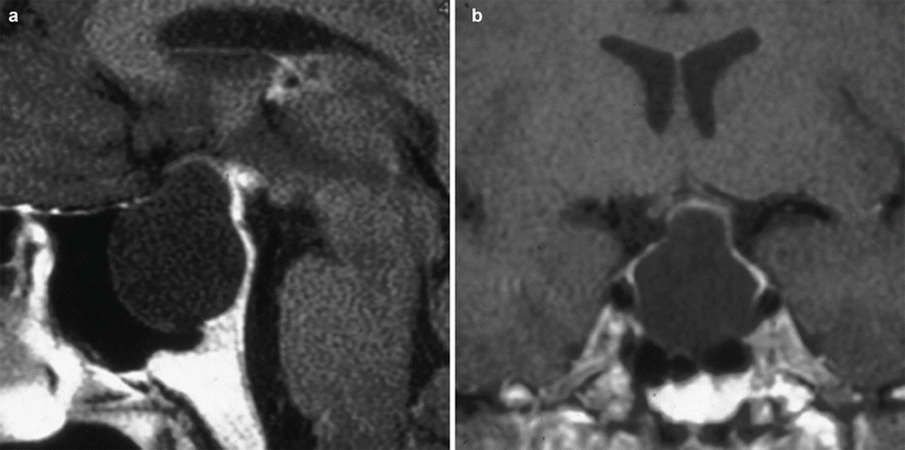
Fig. 25.2
Arachnoid cyst. (a) Sagittal T1-weighted post-gadolinium image. (b) Coronal T1-weighted post-gadolinium image. A nonenhancing lesion in the sella is present with suprasellar extension and remodeling of the sellar floor. The signal intensity of the lesion is similar to the CSF. The optic chiasm is mildly elevated by the mass
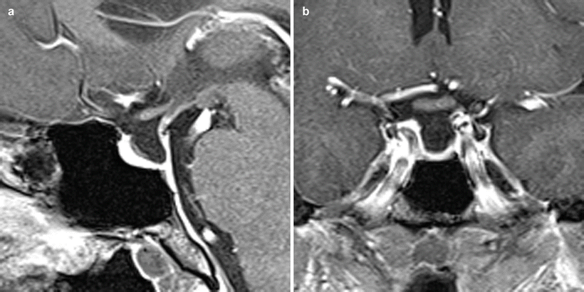
Fig. 25.3
Arachnoid cyst. (a) Sagittal T1-weighted post-gadolinium image. (b) Coronal T1-weighted post-gadolinium image. There is posterior displacement of the pituitary stalk; the depressed appearance of the pituitary gland suggests the presence of a collection with signal intensity similar to that of the CSF. The wall of the cystic collection is not visualized on these images
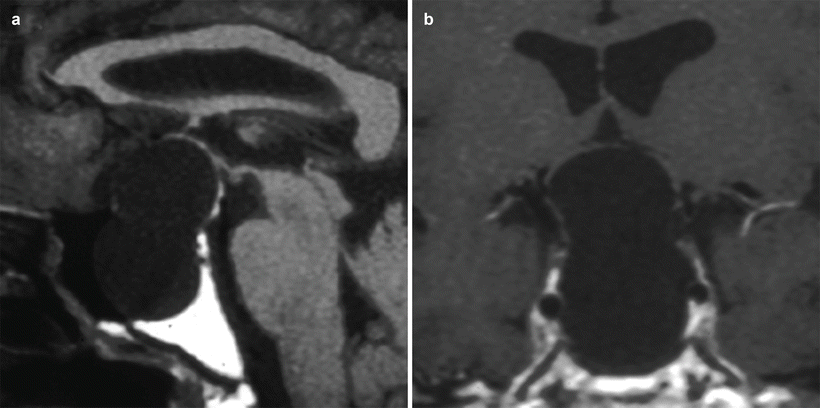
Fig. 25.4
Arachnoid cyst. (a) Sagittal T1-weighted post-gadolinium image. (b) Coronal T1-weighted post-gadolinium image. There is a nonenhancing lesion in the sella, with suprasellar extension and remodeling of the sellar floor. The signal intensity of the lesion is similar to the CSF. The optic chiasm is elevated by the mass
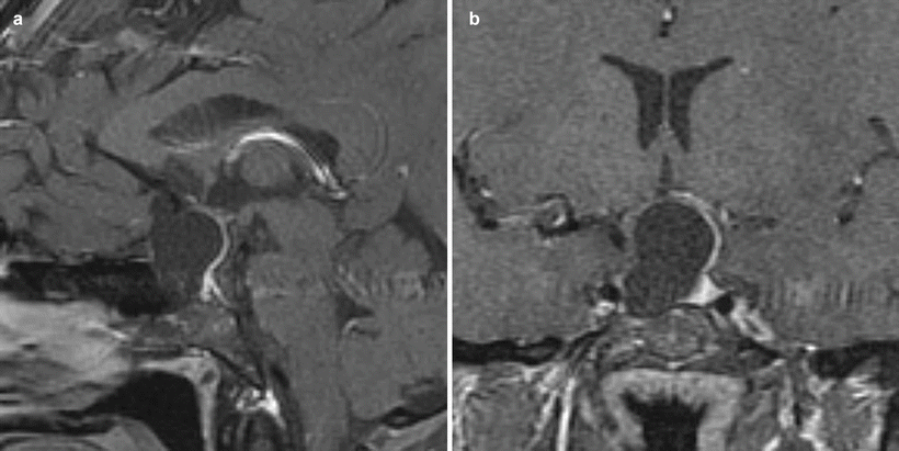
Fig. 25.5
Arachnoid cyst. (a) Sagittal T1-weighted post-gadolinium image. (b) Coronal T1-weighted post-gadolinium image. A nonenhancing lesion is located in the right aspect of the sella, with suprasellar extension and remodeling of the sellar floor. The signal intensity of the lesion is similar to the CSF. The optic chiasm is elevated by the mass
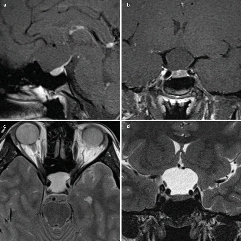
Fig. 25.6
Arachnoid cyst. (a) Sagittal T1-weighted precontrast image. (b) Coronal T1-weighted precontrast image. (c) Axial T2-weighted image. (d) Coronal T2-weighted image. A nonenhancing suprasellar collection displays signal intensity similar to that of the CSF. There is downward effacement of the sellar contents by the collection. The A1 segments of both anterior cerebral arteries are elevated by the collection
25.3 Histopathology
Pathophysiology: Normally, no arachnoid layer is located below the level of the diaphragma sellae. Arachnoid cysts are thought to develop in this region secondary to a congenitally wide aperture in the diaphragma sellae, allowing arachnoid and cerebrospinal fluid (CSF) to enter the sella turcica via a ball-valve effect. Ultimately, the arachnoid membrane apposes and the CSF space no longer communicates, forming a sequestered (noncommunicating) cyst with an arachnoid lining, as opposed to an empty sella condition. (See “Empty Sella Syndrome” in Chap. 66.)
The structure of an arachnoid cyst wall is similar to that of normal arachnoid membrane.
The inner surface of the cyst wall is composed of arachnoid cells with slender processes and large amounts of extracellular space; microvilli are not found (Fig. 25.7) [8].
Immunostaining of the cyst wall can be used to differentiate arachnoid cysts from epithelial cysts, because the arachnoid cysts demonstrate negative immunoreactivity for glial fibrillary acidic protein (GFAP), S-100 protein, prealbumin, and carcinoembryonic antigen (CEA) [9].
Stay updated, free articles. Join our Telegram channel

Full access? Get Clinical Tree


