Fig. 12.1
Growth hormone (GH)–secreting adenoma. (a) Axial noncontrast CT scan with bony windowing. (b) Intraoperative endoscopic photo. The CT scan shows the large caliber and prominence of the internal carotid arteries, which demonstrate ectasia and tortuosity with extension into the sella (white arrows). Adapted from Zada et al. with permission [13]
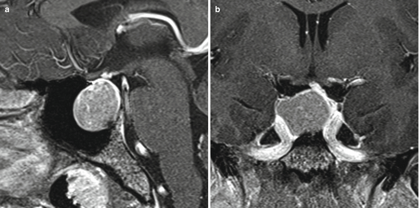
Fig. 12.2
GH adenoma. (a) Sagittal T1-weighted post-gadolinium image. (b) Coronal T1-weighted post-gadolinium image. An expansile sellar/suprasellar mass abuts the right cavernous internal carotid artery, and there is remodeling of the sellar floor. The pituitary stalk is elevated and deviated to the left
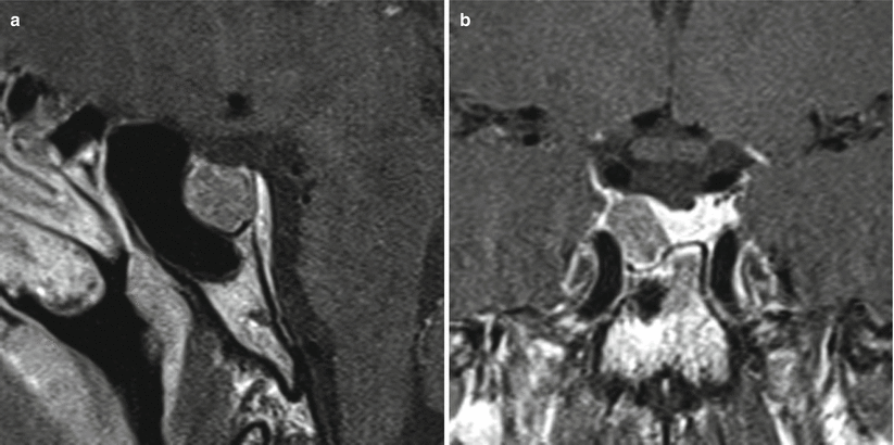
Fig. 12.3
GH adenoma. (a) Sagittal T1-weighted post-gadolinium image. (b) Coronal T1-weighted post-gadolinium image. A hypoenhancing mass presents in the right aspect of the sella abutting the right cavernous internal carotid artery without encasement. There is erosion of the right sellar floor
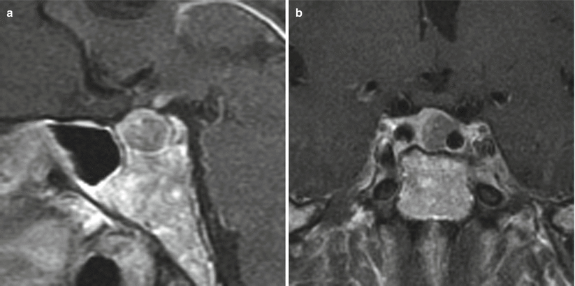
Fig. 12.4
GH adenoma. (a) Sagittal T1-weighted post-gadolinium image. (b) Coronal T1-weighted post-gadolinium image. A hypoenhancing mass is located in the left aspect of the sella, abutting the left cavernous internal carotid artery without encasement
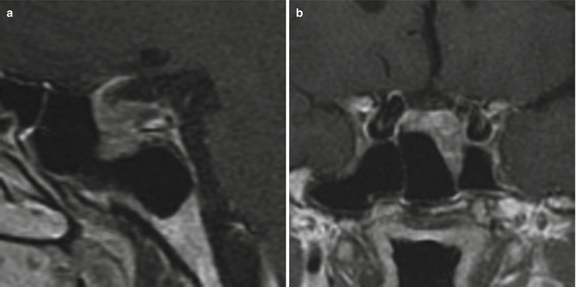
Fig. 12.5
GH adenoma. (a) Sagittal T1-weighted post-gadolinium image. (b) Coronal T1-weighted post-gadolinium image. Within the sella there is a heterogeneous, predominantly hypoenhancing mass causing erosion of the sellar floor. The mass contacts the left cavernous internal carotid artery without evidence of invasion
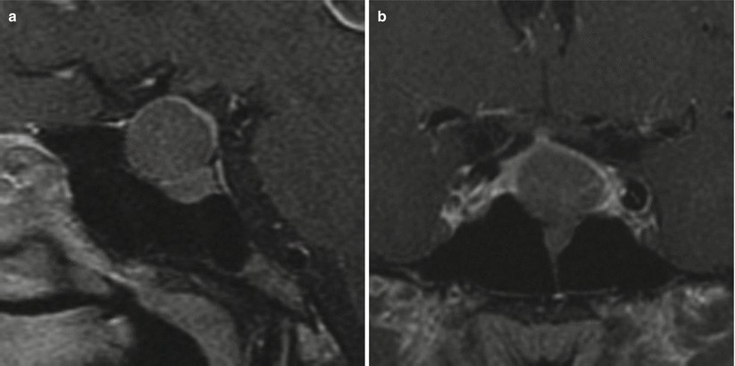
Fig. 12.6
GH adenoma. (a) Sagittal T1-weighted post-gadolinium image. (b) Coronal T1-weighted post-gadolinium image. There is a hypoenhancing mass in the sella causing erosion of the sellar floor and suprasellar extension without contacting the optic chiasm
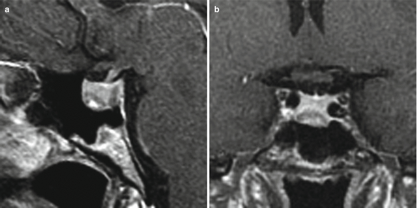
Fig. 12.7
GH adenoma. (a) Sagittal T1-weighted post-gadolinium image. (b) Coronal T1-weighted post-gadolinium image. A small, hypoenhancing mass is seen in the inferior anterior aspect of the sella, causing mild erosion of the sellar floor
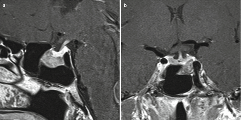
Fig. 12.8
GH adenoma. (a) Sagittal T1-weighted post-gadolinium image. (b) Coronal T1-weighted post-gadolinium image. There is a hypoenhancing mass in the left inferior aspect of the sella, causing erosion of the sellar floor. The mass contacts the left cavernous internal carotid artery without evidence of invasion of the left cavernous sinus
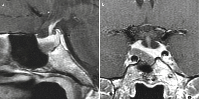
Fig. 12.9
GH adenoma. (a) Sagittal T1-weighted post-gadolinium image. (b) Coronal T1-weighted post-gadolinium image. There is a hypoenhancing mass located in the right inferior aspect of the sella, causing erosion of the sellar floor. The mass abuts the right cavernous internal carotid artery without evidence of encasement
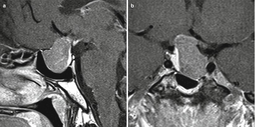
Fig. 12.10
GH adenoma. (a) Sagittal T1-weighted post-gadolinium image. (b) Coronal T1-weighted post-gadolinium image. There is a heterogeneously enhancing sellar mass with suprasellar extension. The mass abuts the left cavernous internal carotid artery without definite invasion of the left cavernous sinus. There is erosion of the left sellar floor. Superiorly, the mass abuts the right hypothalamus. Normal pituitary glandular tissue is seen along the right aspect of the sella
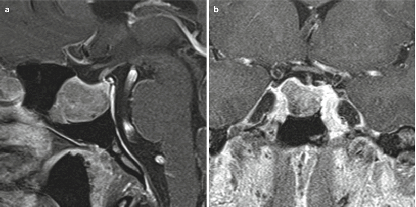
Fig. 12.11
GH adenoma. (a) Sagittal T1-weighted post-gadolinium image. (b) Coronal T1-weighted post-gadolinium image. There is a hypoenhancing sellar mass with erosion of the sellar floor. The mass abuts the bilateral cavernous internal carotid arteries without encasement
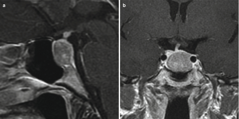
Fig. 12.12
GH adenoma. (a) Sagittal T1-weighted post-gadolinium image. (b) Coronal T1-weighted post-gadolinium image. A hypoenhancing sellar mass erodes the sellar floor and abuts the left cavernous internal carotid artery without encasement. The pituitary stalk is mildly deviated to the right, with normal gland flattened in the superior right aspect of the sella
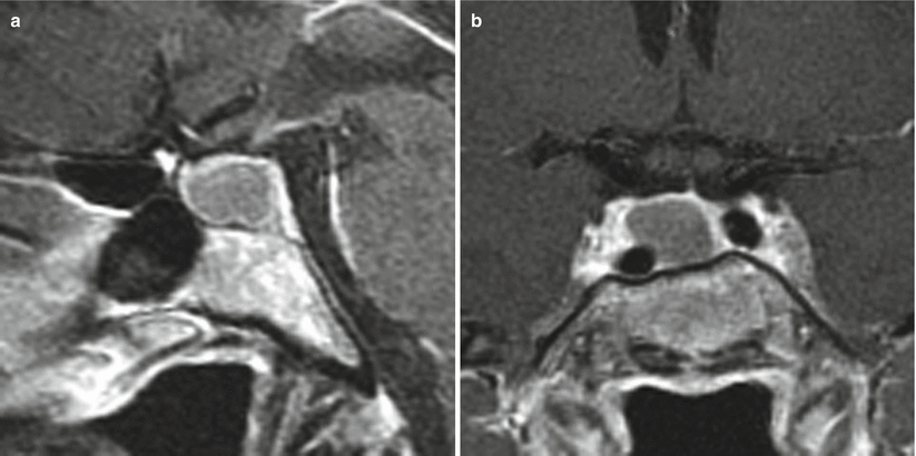
Fig. 12.13
GH adenoma. (a) Sagittal T1-weighted post-gadolinium image. (b) Coronal T1-weighted post-gadolinium image. There is a hypoenhancing sellar mass in the right aspect of the sella, contacting the right cavernous internal carotid artery without encasement
12.2.3 Histopathology
Approximately two thirds of GH adenomas are densely granulated adenomas; the other third are sparsely granulated [2, 14].
Densely granulated GH adenomas are characterized by medium- to large-sized cells with slight pleomorphism and a strong, diffuse staining pattern for GH (Fig. 12.14).
Sparsely granulated GH adenomas are frequently pleomorphic, with fibrous bodies, a region of perinuclear clearing, and typically weaker immunoreactivity for GH stain (Fig. 12.15) [2]. Fibrous bodies in the cytoplasm can be stained with CAM 5.2 [14].
Dural invasion has been noted in over 50 % of GH adenomas [2].
Associated tumor subtypes are those composed of both GH and PRL cells, the mammosomatotroph cell adenoma, and the acidophil stem cell adenoma.
GH/PRL tumors may be more primitive subtypes; they are more likely to be atypical adenomas than pure GH tumors [15].
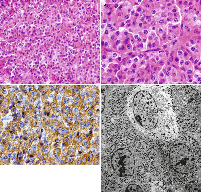
Fig. 12.14
GH adenoma. (a, b) Densely granulated GH cell adenomas show large cells with eosinophilic, granular cytoplasm and a central nucleus with prominent nucleoli. (c) On immunohistochemistry, the tumor shows intense and diffuse staining for GH. (d) Densely granulated GH cell adenoma exhibits well-developed organelles and abundant, large secretory granules at the ultrastructural level
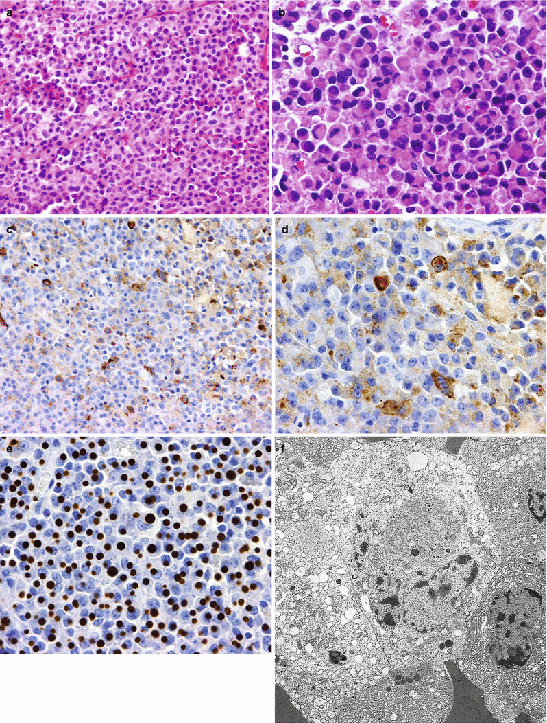
Fig. 12.15
GH adenoma. (a) Sparsely granulated GH cell adenomas have a more chromophobic appearance of the cells, with slight granular cytoplasm. (b) Characteristically, the tumors have a paranuclear eosinophilic inclusion called “fibrous bodies.” (c, d) Immunohistochemistry for GH shows more focal staining within the cell that is less prominent than in densely granulated adenomas. (e) Cytokeratin (CAM 5.2) histochemistry highlights the paranuclear “fibrous body” seen in sparsely granulated GH adenomas. (f) At ultrastructure, sparsely granulated GH adenomas display sparse secretory granules and the characteristic “fibrous bodies”
12.3 Clinical Management
The overall goal of multimodal treatment for acromegaly is the reduction of tumor mass effect and biochemical normalization of IGF-1 and GH levels [8, 16].
Stay updated, free articles. Join our Telegram channel

Full access? Get Clinical Tree


