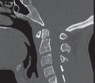
46 What causes occipitoatlantal dislocations?1 Abrupt and violent distraction forces able to displace the occipital condyle from the atlas What is the mechanism of injury in fractures of the occipital condyle? High-energy impact trauma with axial compression with or without basilar skull fracture and ligamentous injury How are occipital condyle fractures classified? The Anderson and Montesano classification system: 1. Type I: impact fracture without ligamentous spread, compound 2. Type II: a fracture of the base of the skull extending through the condyle and communicating with the foramen magnum 3. Type III: fracture of the condyle with incompetence of the ligamentous structure and avulsion of fractured portion of the condyle What fractures of the occipital condyle are always considered unstable?2 Type III What are the possible patterns of fracture of the C1 ring? 1. Isolated posterior arch fracture 2. Anterior arch fracture 3. Comminuted fracture of the lateral masses 4. Burst fracture (Jefferson) What is the mechanism behind an isolated fracture of the posterior arch of C1?3,4 A forced hyperextension of the skull and cervical spine causes compression of the posterior arch that is caught in the middle of two more robust systems (occiput and C2). What is the mechanism behind a Jefferson fracture? • An axial compression applied to the skull, as in patients who suffered a fall from a height. • The force passes through the condyles and spreads outward, causing the ring to possibly break at four different points (in front and behind each lateral mass). What is the most common fracture of the atlas? The isolated posterior arch fracture, accounting for two thirds of all atlas fractures Are fractures of the atlas stable? Fractures of the atlas are considered unstable if the transverse ligament is severely damaged, allowing for lateral mass displacement of C1 over C2. Which fractures are suitable for intervention?5 If this lateral mass displacement is greater than 7 mm, the fracture requires surgical treatment. What is meant by the term atlantoaxial instability and what is the mechanism of injury? • It refers to injuries of the transverse or the alar ligament or both, in the presence or absence of fractures to the atlas or C2. • Several mechanisms of injury can account for this instability, most commonly including flexion and rotation. How are injuries of the transverse ligament classified? Dickman et al divided these injuries into four types6: IA: midsubstance tear (ligamentous only) IB: lateral tear (at the periosteal attachment) IIA: ligamentous incompetence due to comminuted fracture of the lateral mass IIB: ligamentous detachment with a small piece of the lateral mass In patients with suspected atlantoaxial instability, what atlantodental interval (ADI) is considered unstable?7 • Greater than 3 mm in adults • Greater than 5 mm in children How is atlantoaxial rotational instability classified? In four types according to the system of Fielding and Hawkins that evaluated the ADI and the degree of subluxation of the lateral mass joints8: Type I: no increase of the ADI with fixed rotation of the lateral masses. Type II: ADI less than 5 mm and one lateral mass joint preserved Type III: asymmetrical lateral mass subluxation and ADI more than 5 mm Type IV: Posterior subluxation and rotation What are the three columns of Denis?9 1. The anterior column a. extends from the anterior half of the vertebral body, and b. includes the anterior longitudinal ligament. 2. The middle column a. includes the posterior half of the vertebral body, b. the posterior longitudinal ligament, and c. the pedicles. 3. The posterior column a. includes the posterior elements, b. the facet joints and capsule, c. the ligamentum flavum, d. the spinous processes, and e. the interspinous ligaments. What do the three columns of Dennis represent?10 • The column of Dennis represents a model for assessment of spinal instability in the thoracolumbar spine: • Injury to two of the three columns represents an instability. What is atlanto-occipital dislocation?11 • It is an injury involving the craniocervical junction. • There is instability of the ligamentous elements between the atlas and the occiput. What is a typical presentation of the patient with atlanto-occipital dislocation? • Patients may present with minimal neurological deficit or exhibit more severe findings such as medullary injury or paraparesis. • Extreme cases present with mortality secondary to hypoxia and respiratory arrest. What age group is typically affected with this condition? • The typical patient is usually a pediatric patient, as the shape of the condyles is flatter in children and their heads are proportionately larger compared with the rest of the body. What are the types of atlanto-occipital dislocation? There are three types: Type I: anterior dislocation Type II: distraction injury Type III: posterior dislocation Discuss one way of evaluating atlanto-occipital dislocation by imaging studies.12 Plain x-rays and a CT scan of the cervical spine and craniocervical junction are obtained, and the Powers ratio is evaluated. What is the Powers ratio? The Powers ratio identifies anterior subluxation and is described as a ratio of BC/OA. BC is the distance from the basion to the midvertical portion of the posterior laminar line of the atlas. OA is the distance from the opisthion to the midvertical portion of the posterior surface of the anterior ring of Atlas. If this ratio is greater than 1, anterior subluxation exists. How it is atlanto-occipital dislocation managed in the acute phase? • The cervical spine is immediately immobilized. • This may be done by using a halo vest. • A hard collar is usually not enough for stabilization. What is the definitive management of atlanto-occipital dislocation? • In several cases posterior fixation with occipital cervical fusion is required. • In mild cases or gray-zone cases, a halo vest may be enough for immobilization while waiting for the ligaments to heal. What is a hangman’s fracture?1 It is a fracture involving the pars articularis of C2 bilaterally, usually caused by a hyperextension injury. What is the mechanism behind hangman’s fracture? Typical causes include hyperextension and axial loading or axial distraction as in judicial hanging Describe a classification of hangman’s fracture The Levine/Effendi classification has been described for hangman’s fracture: • Type 1: • Pars fracture with less than 3-mm subluxation of C2 on C3, and • No angulation • Type 1A: • The fracture may pass through the foramen transversarium as the fracture lines are not parallel, and • The vertebral body may appear elongated (atypical type). • Type 2: • Fracture through the pars with disruption of the C2–3 disk space, and • Subluxation of greater than 3 mm. • Type 2A: • Oblique fracture with less subluxation (less than 3 mm), • But angulation greater than 15 degrees. • This fracture is typically unstable. • Type 3: • A type 2 fracture with bilateral C2–3 facet disruption with subluxation or locked facets. • This is a highly unstable fracture that requires internal reduction. How are hangman’s fractures managed initially?2 Initial management as well as type 1 management involving the use of external orthosis such as a halo or collar What are the indications for surgery? • Surgical fixation is typically done for type 2 fractures with • more severe angulation (>15 degrees), • severe subluxation (>5 mm), or • disruption of the disk space. • Surgery may also be considered in cases that have failed external immobilization with inadequate alignment. What are the surgical options to treat hangman’s fractures? • C2–3 anterior cervical diskectomy and fusion • C1–3 lateral mass fixation with rods and screws • C1–3 wiring and fusion • A combination of the above techniques with anterior and posterior fixation and fusion • Placement of a screw through the fracture fragments (C2 pedicle screw). This technique typically requires neuronavigational or biplanar radiography. Described the classification of odontoid fractures.1 There are three types of fractures: Type I is a fracture through the tip of the odontoid process. It may be unstable in certain cases. Type II is a fracture through the base of the odontoid process. It is usually unstable Type III is a fracture through the body of the C2 vertebra. It may involve the articulating facet and is usually stable. Fig. 46.1 Sagittal reconstructed CT scan of the cervical spine demonstrating C2 odontoid type II fracture with minimal displacement. This fracture is amenable to placement of an odontoid screw.
Spinal Column Trauma
46.1 Classification of Fractures and Mechanisms of Injury
46.2 Atlanto-Occipital Dislocation
46.3 Hangman’s Fracture
46.4 Odontoid Fractures

Spinal Column Trauma
Only gold members can continue reading. Log In or Register to continue

Full access? Get Clinical Tree


