FIGURE 45.1 General arrangement of the autonomic nervous system. The sympathetic components are shown in red, the parasympathetic component in blue. (From Snell R. Clinical Neuroanatomy. 7th ed. Philadelphia: Wolters Kluwer/Lippincott Williams & Wilkins, 2009, with permission.)
THE PERIPHERAL AUTONOMIC NERVOUS SYSTEM
The parasympathetic division is composed of the general visceral efferent fibers of cranial nerves III, VII, IX, X, and bulbar portion of XI (the cranial outflow), together with fibers arising in the S2-S4 segments of the spinal cord (the sacral outflow). The parts of the parasympathetic division are widely separated, but because of anatomic characteristics, similarity in function, and similar pharmacologic responses, they are classified as parts of one system rather than as separate divisions. The parasympathetic nerves have long preganglionic fibers that end in peripheral ganglia near or in the viscera they supply, and short postganglionic fibers that arise in proximity to or within the viscus innervated. One preganglionic fiber usually synapses with only one postganglionic neuron.
The anatomy of the cranial portion of the parasympathetic division is discussed with the individual cranial nerves. In brief, it consists of the Edinger-Westphal nucleus, the superior and inferior salivatory nuclei, the dorsal motor nucleus of the vagus, and neurons in the vicinity of the nucleus ambiguus. The sacral parasympathetic fibers arise from cells in the intermediolateral cell column at the S2-S4 levels of the sacral spinal cord, travel through the sacral nerves, and are collected into the pelvic splanchnic nerves (nervi erigentes), which proceed to the pelvic plexuses and their branches. Some postganglionic fibers may travel from these plexuses to the pelvic viscera, but most preganglionic fibers continue to small ganglia in or near the viscera, from where postganglionic fibers supply the bladder, descending colon, rectum, anus, and genitalia. The greatest parasympathetic outflow is via the vagus nerves. Peripheral parasympathetic ganglia include the ciliary, otic, submandibular, and sphenopalatine (Figure 45.2).
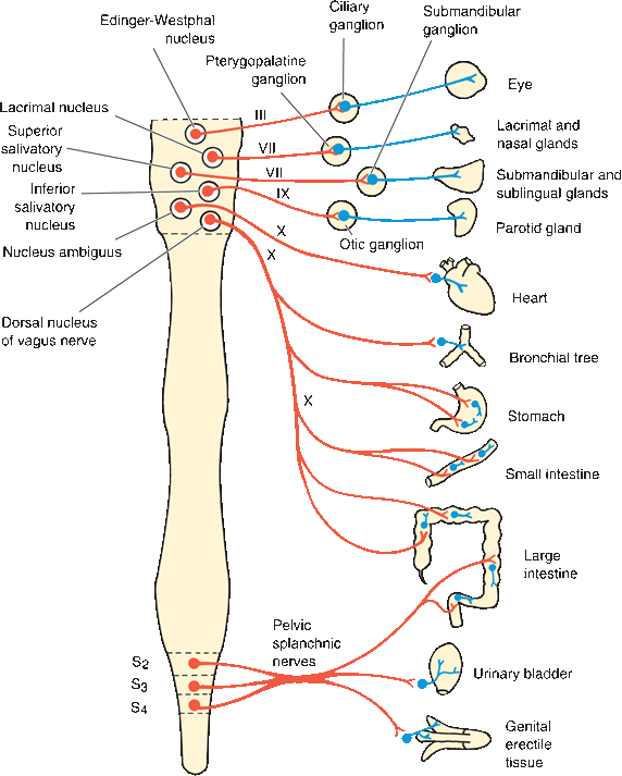
FIGURE 45.2 The parasympathetic division. Preganglionic neurons are red, and postganglionic neurons are blue. (From Kiernan JA. Barr’s the Human Nervous System: An Anatomical Viewpoint. 9th ed. Philadelphia: Wolters Kluwer/Lippincott Williams & Wilkins, 2009, with permission.)
The sympathetic division is composed of preganglionic fibers that arise from cells in the intermediolateral columns from the T1 to the L3 segments of the spinal cord. The fibers exit through the ventral roots of the corresponding segmental nerves (Figure 24.3). These fibers terminate in the paravertebral ganglionic chain, the prevertebral plexuses and collateral ganglia, or occasionally the terminal ganglia (Figure 45.3). Postganglionic fibers go to the viscera. The sympathetic preganglionic fibers are typically short and terminate on ganglia some distance from the viscera they supply, with long postganglionic fibers that travel from the ganglia to the viscera. One preganglionic fiber may synapse with many postganglionic neurons.
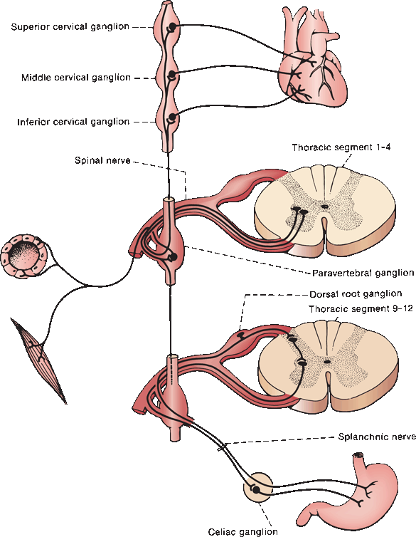
FIGURE 45.3 The sympathetic outflow, showing connections with the paravertebral ganglionic chain, splanchnic nerves, and collateral ganglia.
The sympathetic ganglia are arranged into two plexuses: paravertebral and prevertebral. The paravertebral ganglia lie alongside the vertebral column; the prevertebral ganglia lie anterior to the vertebral column. The prevertebral ganglia innervate the viscera of the abdomen and pelvis. The paravertebral sympathetic chain consists of two elongated plexuses, each composed of a series of ganglia that are segmentally arranged and bound together by ascending and descending nerve fibers. The sympathetic trunks have from 22 to 24 ganglia and extend from the level of C2 to the coccyx. There are 3 cervical, 10 to 12 thoracic, 4 lumbar, and 4 to 5 sacral ganglia. The chains usually join at the level of the coccyx in an unpaired coccygeal ganglion (ganglion impar). Preganglionic fibers leave the spinal cord through the anterior root and mixed spinal nerve to reach the anterior primary ramus, and then exit as finely myelinated fibers (white rami communicantes) to enter the ganglionic chain. They may synapse immediately or ascend or descend before synapsing. The postganglionic fibers return to the anterior primary ramus as unmyelinated fibers (gray rami communicantes). The T1-T3 segments innervate the head and neck, the T3-T11 segments innervate the upper extremities and viscera in the thorax and abdomen, and the T12-L2 segments innervate the lower extremities and pelvic viscera.
The cervical portion of the sympathetic chain consists of the superior, middle, and inferior cervical ganglia. These innervate structures within the head, upper extremities, and thorax. The superior cervical ganglion, the largest, lies opposite the C2-C3 vertebrae and behind the internal carotid artery. It is primarily supplied by the first two thoracic segments. The internal carotid nerve, a direct continuation of the superior cervical ganglion, gives rise to postganglionic filaments that supply the internal carotid and terminate as the internal carotid and cavernous plexuses. Anterior branches from the ganglion form plexuses around the middle meningeal and external carotid and maxillary arteries. The sympathetic innervation of the ciliary ganglia travels through the long ciliary nerves from the cavernous plexus. The sphenopalatine ganglion is supplied by the internal carotid plexus through the deep petrosal and vidian nerves. The otic ganglion receives its sympathetic innervation from the plexus around the middle meningeal artery, and the submaxillary ganglion from that around the external maxillary artery. There are other connections from the superior cervical ganglion to other cranial nerves and the upper four cervical nerves, the pharyngeal plexus, the carotid sinus and body, the heart, and the superior cardiac nerves. The middle cervical ganglion communicates with the fifth and sixth cervical nerves to begin the middle cardiac nerve and sends other branches to the thyroid gland. The inferior cervical ganglion communicates with the seventh and eighth cervical nerves to form the inferior cardiac nerve and nerves to the blood vessels.
The paravertebral ganglia provide long unmyelinated axons to all sympathetically innervated tissues and organs except those in the abdomen, pelvis, and perineum. The superior cervical ganglion (T1-T2) provides pupillodilator and sudomotor fibers to the face. The stellate ganglion (T2-T6) innervates the upper limb through branches of the brachial plexus, and the lumbar sympathetic ganglia (T9-L1) innervate the lower limb through branches of the lumbosacral plexus. The postganglionic sympathetic fibers join the peripheral somatic nerves via the gray rami communicantes, and thus, their distribution is similar to that of the corresponding somatic nerve.
The thoracic portion of the sympathetic trunk rests against the heads of the ribs. Occasionally, the first thoracic ganglion is blended with the inferior cervical ganglion to form the stellate ganglion. The stellate ganglion receives preganglionic fibers from the T2-T6 levels, and its postganglionic fibers are distributed with the nerves of the brachial plexus to provide autonomic innervation to the upper extremity. The sympathetic fibers traveling in somatic nerves innervate vasomotor, sudomotor, and pilomotor structures in the distribution of the nerve in which they are carried.
The upper five ganglia provide branches to the cardiac and pulmonary plexuses. The abdominal portion of the sympathetic trunk is situated in front of the vertebral column along the medial margin of the psoas major muscle, and the pelvic portion is in front of the sacrum. All of these ganglia send gray rami communicantes to the corresponding spinal nerves and many branches to the various plexuses and collateral ganglia. The postganglionic fibers terminate on blood vessels, sweat glands, and other smooth muscle and glandular structures.
Branches of the lower seven thoracic ganglia unite to form the three splanchnic nerves that penetrate the diaphragm and supply the abdomen and the pelvic viscera. These branches are white in color and primarily carry preganglionic fibers that pass through the ganglia without synapsing and terminate in the prevertebral plexuses or the collateral ganglia. The greater splanchnic nerve is formed by branches of the fifth through the 9 th or 10th thoracic ganglia; it terminates in the celiac ganglion. The lesser splanchnic nerve is formed by branches of the 9th, 10th, and sometimes the 11th thoracic ganglia; it ends in the aorticorenal ganglion. The lower splanchnic nerve arises from the last thoracic ganglion; it ends in the renal plexus.
Within the thoracic, abdominal, and pelvic cavities are aggregations of nerves and ganglia known as the prevertebral plexuses and their collateral ganglia. These are composed of both parasympathetic and sympathetic fibers. The parasympathetic fibers are preganglionic and may synapse in the plexuses or go through without synapse to terminal ganglia. The sympathetic fibers, mainly from the splanchnic nerves, usually synapse in the plexuses. From these plexuses, branches are given off to the abdominal and pelvic viscera. The cardiac plexus is supplied by the cardiac branches of the vagus nerves and the cardiac nerves arising from the cervical and upper thoracic sympathetic ganglia. The cardiac plexus also communicates with the pulmonary and the esophageal plexuses, all supplied by the vagus nerve as well as the thoracic sympathetic ganglia.
The celiac plexus is the largest of the three sympathetic plexuses and innervates all the abdominal viscera except for the descending colon. The thoracic splanchnic nerves, carrying preganglionic fibers from the T5-T12 levels, perforate the diaphragm and form the celiac plexus, which lies in the abdomen at the level of the upper part of the first lumbar vertebra, behind the stomach and omental bursa, in front of the diaphragm and abdominal aorta, and between the adrenal glands. It is composed of the two celiac ganglia that are supplied by the greater splanchnic nerves and filaments from the right vagus nerve, and the aorticorenal ganglia, which receive the lesser splanchnic nerves. Other plexuses arise from or are connected with the celiac plexus, including phrenic, hepatic, splenic, and others. The superior (anterior) gastric plexus and the hepatic plexus also receive branches from the left vagus nerve. The renal and inferior mesenteric plexuses and their branches are also supplied by the lowest splanchnic nerve.
The hypogastric plexus is located in front of the last lumbar vertebra and the promontory of the sacrum, between the two common iliac arteries, and is formed by the union of many elements from the aortic plexus and the lumbar sympathetic chain, together with some fibers from the inferior mesenteric plexus. It is divided into the two pelvic plexuses formed by fibers from the hypogastric plexus; preganglionic sympathetic fibers from the second, third, and fourth sacral nerves; and a few filaments from the sacral sympathetic ganglia. Branches are distributed to the pelvic viscera and the internal and external genitalia through the middle hemorrhoidal, vesical, prostatic, vaginal, and uterine plexuses.
The enteric nervous system consists of intrinsics and extrinsic components. The intrinsic component consists of Meissner’s submucosal and Auerbach’s myenteric plexuses. The extrinsic component consists of preganglionic sympathetic, from prevertebral ganglia, and parasympathetic, from the dorsal motor nucleus of the vagus and the sacral parasympathetic centers, inputs that control peristalsis and secretion.
Autonomic Afferents
General visceral afferent fibers convey both conscious and unconscious sensations from the viscera, and are involved in autonomic reflexes. Small myelinated and unmyelinated fibers carry impulses from visceral receptors to cell bodies in the dorsal root and cranial nerve ganglia. The visceral afferents that enter the spinal cord synapse on neurons in the dorsal horn and intermediolateral gray column. Centrally, sensation from the viscera travels mainly in the spinothalamic and spinoreticular tracts, but some visceral afferents—especially those related to bowel and bladder control—are carried in the posterior columns. After a synapse in the thalamus, visceral sensory fibers project to areas of the cortex involved in autonomic function. Afferent autonomic fibers in the vagus nerve synapse in the nodose ganglion, those in the glossopharyngeal nerve in the petrosal ganglion. The vagal afferents transmit impulses from the heart, great vessels, lungs, and GI tract; the glossopharyngeal afferents convey information from the carotid sinus. These afferents synapse in the nucleus of the solitary tract (NST) and are involved in autonomic reflexes as well as such functions as coughing and swallowing.
Neurotransmitters
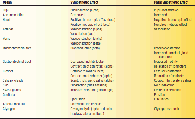
Acetylcholine is the neurotransmitter at sympathetic and parasympathetic preganglionic neurons, and at postganglionic parasympathetic neurons. Norepinephrine is the primary postganglionic sympathetic neurotransmitter, except at sweat glands, which are cholinergic. There are two subtypes of acetylcholine receptor: nicotinic and muscarinic. Most of the postganglionic acetylcholine receptors are muscarinic. They mediate the cardiac effects and cause pupillary constriction, lacrimal and salivary secretion, bronchoconstriction, and erection. They also stimulate GI tract motility and cause evacuation of the bladder and rectum. There are two main subtypes of adrenergic receptors: alpha and beta. The alpha-adrenergic receptors mediate pupillary dilatation, vasoconstriction, and ejaculation, and they also control the internal sphincters of the bladder and rectum. Beta-adrenergic receptors control the heart, cause vasodilation and bronchial dilatation, and mediate metabolic effects. Some postganglionic sympathetic neurons also utilize adenosine triphosphate and neuropeptide Y, and some postganglionic parasympathetic endings may use vasoactive intestinal polypeptide or nitric oxide.
The Physiology of the Peripheral Autonomic Nervous System
The ANS governs the activities of cardiac and smooth muscle, including the smooth muscle of the blood vessels and the functions of most glandular structures. It regulates such important functions as respiration, circulation, digestion, temperature adjustment, and metabolism—all vital to normal existence—and combats forces acting from within or without that would tend to cause undesirable changes in the normal function of the body. By homeostasis, the constancy of the internal environment of the body and the uniformity and stability of the organism are maintained.
The sympathetic division supplies all parts of the body. Its functions are catabolic and directed toward the utilization of energy. It prepares the organism for combat or escape (fight-or-flight response). It acts whenever rapid adjustment to the environment is required. It accelerates the heart, dilates the coronary vessels, increases the arterial blood pressure (BP), empties the blood reservoirs, dilates the bronchi, liberates glucose, and inhibits GI activity. It is an emergency protective mechanism that is called into action under emotional stress and causes the individual to react strongly to stimuli of rage and fear. The parasympathetic division supplies special structures, such as the pupils, salivary glands, heart, lungs, GI tract, bladder, and portions of the genital system. In certain parasympathetic functions, as in bladder, rectal, and genital activity, contraction of striated muscles are closely integrated with those of smooth muscle. The parasympathetic division conserves energy. It controls anabolic, excretory, and reproductive functions, and conserves and restores bodily resources and energy.
The viscera receive a dual autonomic supply, both sympathetic and parasympathetic. In general, these two divisions are antagonistic and reciprocal in their functions, but there are exceptions. Table 45.1 compares the functions of the two divisions in the innervation of various effector organs.
TABLE 45.1 Effects of Sympathetic and Parasympathetic Systems on Various Effector Organs
THE CENTRAL REGULATION OF AUTONOMIC FUNCTION
The peripheral ANS is under the control of higher centers in the cerebral cortex, especially the amygdala, hypothalamus, basal forebrain, ventral striatum, brainstem, and spinal cord that regulate and influence the function of its peripheral components. The centers in the CNS that are involved in autonomic function are referred to as the central autonomic network. The neurons of the central autonomic network are interconnected and make up a functional unit. The most important of these centers is the hypothalamus.
The Hypothalamus
The hypothalamus (Figure 45.4) is part of the ventral diencephalon, lying just below the thalamus and above the pituitary gland. The entire area measures only about 14 × 18 × 20 mm and weighs only 4 g. It forms most of the floor and part of the lateral wall of the third ventricle, extending from the level of the chiasm to the interpeduncular fossa. From a strictly anatomic point of view, it includes the optic chiasm, neurohypophysis (posterior pituitary), infundibulum, pars supraoptica, tuber cinereum, and mammillary bodies, but from a physiologic point of view, the first three structures are not included. The supraoptic nucleus is located just above the optic chiasm. The mammillary bodies are a pair of small spherical gray matter masses that lie in the interpeduncular fossa rostral to the posterior perforated substance and contain the mammillary nuclei; they form the caudal portion of the hypothalamus. Beneath the hypothalamus, a hollow, conical process, the infundibulum, or pituitary stalk, projects downward and forward and is attached to the posterior lobe of the hypophysis. The infundibulum contains the supraopticohypophyseal and tuberohypophyseal tracts. The tuber cinereum is a prominence that lies between the mammillary bodies and the infundibulum.
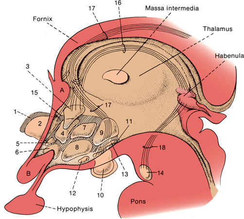
FIGURE 45.4 Diagrammatic sketch of the human hypothalamus. A. Anterior commissure. B. Optic nerve. (1). Lateral preoptic area, permeated by the median forebrain bundle. (2). Medial preoptic area. (3). Paraventricular nucleus. (4). Anterior hypothalamic area. (5). Suprachiasmatic nucleus. (6). Supraoptic nucleus. (7). Dorsomedial hypothalamic nucleus. (8). Ventromedial hypothalamic nucleus. (9). Posterior hypothalamic area. (10). Medial mammillary nucleus. (11). Lateral mammillary nucleus. (12). Premammillary area. (13). Supramammillary area. (14). Interpeduncular nucleus. (15). Lateral hypothalamic area. (16). Stria habenularis. (17). Fornix. (78). Habenulopeduncular tract
Related posts:

Stay updated, free articles. Join our Telegram channel

Full access? Get Clinical Tree


