TABLE 38.2 Method of Recording the Commonly Tested Muscle Stretch Reflexes
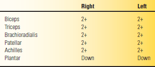
Grades 0 to 4+ (see text) used for all plantar reflex, which is down (normal), absent (0), equivocal (+/–) or up (abnormal). Other reflexes may be added and charted as needed.
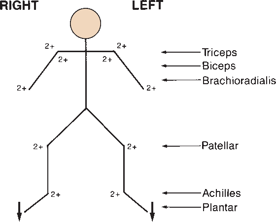
FIGURE 38.1 Alternate method of recording the commonly tested muscle stretch reflexes. For grading, see text and Table 38.2.
In some patients, DTRs may be markedly diminished, or even apparently absent, although there is no other evidence of nervous system disease. Under such circumstances, reinforcement techniques are often useful. Reflex reinforcement probably involves supraspinal, fusimotor, and long-loop mechanisms. A reflex can be reinforced or brought out using several methods. In the Jendrassik maneuver, the patient attempts to pull the hands apart with the fingers flexed and hooked together, palms facing, as the tendon is percussed (Figure 38.2). The effect is very brief, lasting only 1 to 6 seconds, and is maximal for only 300 milliseconds. The Jendrassik maneuver is obviously only useful for lower-extremity reflexes. Other techniques include having the patient clench one or both fists, firmly grasp the arm of the chair, side of the bed, or the arm of the examiner. Reinforcement may also be carried out by having the patient look at the ceiling, grit the teeth, cough, squeeze the knees together, take a deep breath, count, read aloud, or repeat verses at the time the reflex is being tested. A sudden loud noise, a painful stimulus elsewhere on the body—such as the pulling of a hair or a bright light flashed in the eyes—may also be a means of reinforcement.
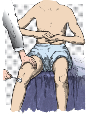
FIGURE 38.2 Method of reinforcing the patellar reflex.
Procedures other than distraction are also helpful in reflex reinforcement. A slight increase in tension of the muscle being tested may reinforce the reflex response. A simple and effective method to reinforce a knee or ankle jerk is to have the patient maintain a slight, steady contraction of the muscle whose tendon is being tested (e.g., slight plantar flexion by pushing the ball of the foot against the floor or the examiner’s hand to reinforce the ankle jerk). The patient may tense the quadriceps by extending the knee slightly against resistance as the knee jerk is being elicited. Reinforcement may increase the amplitude of a sluggish reflex or bring out a latent reflex not otherwise obtainable. Reflexes that are normal on reinforcement, even though not present without reinforcement, may be considered normal. Slight muscle contraction due to inability to relax may be one reason for the slightly hyperactive reflexes often seen in patients who are tense or anxious.
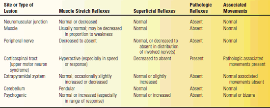
The DTRs are instrumental in the evaluation of weakness. Under most circumstances, weakness accompanied by hyporeflexia is of lower motor neuron origin, and weakness accompanied by hyperre-flexia of upper motor neuron origin. The presence of pathologic reflexes (see Chapter 40) and abnormalities of associated movements (see Chapter 42) are also helpful in the differential diagnosis (Table 38.3). The following sections discuss the upper-extremity, trunk, and lower-extremity reflexes. The masseter or mandibular reflex (jaw jerk) is covered in Chapter 15.
TABLE 38.3 Reflex Patterns with Different Neurologic Disorders
THE UPPER-EXTREMITY REFLEXES
The biceps, triceps, brachioradialis, and finger flexor reflexes are the most important upper-extremity reflexes.
The Biceps Reflex
With the arm relaxed and the forearm slight pronated and midway between flexion and extension, the examiner places the palmar surface of her extended thumb or finger on the patient’s biceps tendon and then strikes the extensor surface with the reflex hammer (Figure 38.3). Pressure on the tendon should be light; too much pressure exerted with the thumb or finger against the tendon makes the reflex much harder to obtain. The hands may lie in the patient’s lap, or the examiner may hold the patient’s arm with the elbow resting in her hand. The major response is a contraction of the biceps muscle with flexion of the elbow. Since the biceps is also a supinator, there is often a certain amount of supination. If the reflex is exaggerated, the reflexogenic zone is increased and the reflex may even be obtained by tapping the clavicle; there may be abnormal spread with accompanying flexion of the wrist and fingers and adduction of the thumb.
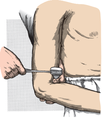
FIGURE 38.3 Method of obtaining the biceps reflex.
The Triceps Reflex
This reflex is elicited by tapping the triceps tendon just above its insertion on the olecranon process of the ulna. The arm is placed midway between flexion and extension and may be rested in the patient’s lap, on her thigh or hip, or on the examiner’s hand (Figure 38.4). The response is contraction of the triceps muscle with extension of the elbow. The most common error in eliciting the triceps jerk is simply too timorous a blow. The paradoxical or inverted triceps jerk consists of flexion of the elbow with percussion of the triceps tendon. This response appears when the afferent arc of the triceps reflex is damaged, as in lesions of the seventh and eighth cervical segments, particularly when there is an element of spasticity, as in cervical spondylosis with radiculomyelopathy.
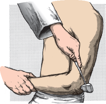
FIGURE 38.4 Method of obtaining the triceps reflex.
The Brachioradialis (Radial Periosteal or Supinator) Reflex
Tapping just above the styloid process of the radius with the forearm in semiflexion and semipronation causes flexion of the elbow, with variable supination (Figure 38.5). The supination is more marked with the forearm extended and pronated, but there is less flexion. The principal muscle involved is the brachio-radialis. The tendon can be percussed not only at its insertion on the lateral aspect of the base of the styloid process of the radius but also at about the junction of the middle and distal thirds of the forearm or at its tendon of origin above the lateral epicondyle of the humerus. The most common error is hitting the muscle belly rather than the tendon. The muscle becomes tendinous at about midforearm. A local contraction can be elicited from any muscle by directly striking the muscle belly. The point in eliciting a DTR is to lengthen the muscle by stretching its tendon. An idiomuscular contraction can be obtained by striking the brachioradialis muscle belly in the proximal third of the forearm; this is not a DTR. If the reflex is exaggerated, there is associated flexion of the wrist and fingers, with adduction of the forearm. When the afferent limb of the reflex is impaired, there may be a twitch of the flexors of the hand and fingers without flexion and supination of the elbow; this is termed inversion of the reflex.
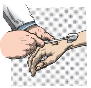
FIGURE 38.5 Method of obtaining the brachioradialis reflex.
The biceps, triceps, and brachioradialis reflexes should be obtained without difficulty in normal individuals. The following upper-extremity reflexes may be elicited only to a slight extent in normal persons. They may become conspicuous when there is hyperreflexia.
The Finger Flexor Reflex (Wartenberg’s Sign)
This is one of several signs attributed to Robert Wartenberg. To elicit the finger flexor reflex, the patient’s hand is in supination, resting on a table or a solid surface, with the fingers slightly flexed. The examiner places her fingers against the patient’s fingers and taps the backs of her own fingers lightly with the reflex hammer (Figure 38.6). The response is flexion of the patient’s fingers and the distal phalanx of the thumb. The reflex may be reinforced by having the patient flex her fingers slightly as the blow is delivered. An alternate technique is for the patient to hold the hand in the air, palm down, and the examiner touching fingers with palm up, with the blow delivered in an upward direction from below. The nerve supply, as in the wrist flexion reflex, is through the median and ulnar nerves (C8-T1). This reflex is difficult for the inexperienced examiner to elicit, and it is often absent in normals. However, Wartenberg considered it one of the most important upper-extremity reflexes. The Hoffmann and Tromner signs, which are pathologic variations of this response, are described in Chapter 40.
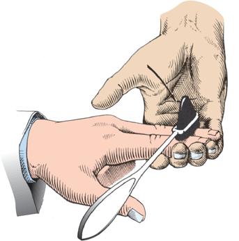
FIGURE 38.6 Method of obtaining the finger flexor reflex.
The Scapulohumeral Reflex
Tapping over the vertebral border of the scapula, either at the tip of its spine or at its base near the inferior angle, causes retraction of the scapula through action of the rhomboid muscles (dorsal scapular nerve, C4-C5). There may be associated elevation of the scapula and adduction and external rotation of the humerus through the trapezius, latissimus dorsi, infraspinatus, and teres minor.
The Deltoid Reflex
Tapping over the insertion of the deltoid muscle at the junction of the upper and middle third of the lateral aspect of the humerus results in slight abduction of the upper arm (axillary nerve, C5-C6).
The Pectoralis Reflex
With the patient’s arm in midposition between abduction and adduction, the examiner places her finger as nearly as possible on the tendon of the pectoralis major muscle near its insertion on the greater tuberosity of the humerus (Figure 38.7). Tapping the finger causes adduction and slight internal rotation of the arm at the shoulder. The contraction of the muscle may be felt but usually not seen in the normal individual. In patients with cervical spondylotic myelopathy, a hyperactive pectoralis reflex indicates spinal cord compression at the C2-C3 and/or C3-C4 levels. This reflex is mediated by the medial and lateral pectoral (anterior thoracic) nerves (C5-T1).
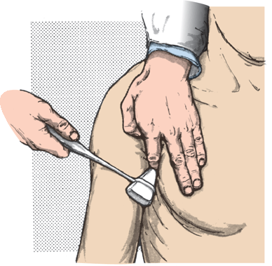
FIGURE 38.7 Method of obtaining the pectoralis reflex.
The Latissimus Dorsi Reflex
With the patient prone and her arm abducted and in slight external rotation, the examiner places her fingers on the tendon of the latissimus dorsi near its insertion in the intertubercular groove of the humerus and taps her finger with the reflex hammer. This produces abduction and slight internal rotation of the shoulder. This reflex is mediated by the thoracodorsal (long subscapular) nerve (C6-C8).
The Clavicle Reflex
In patients with upper-extremity hyperreflexia, a tap over the lateral aspect of the clavicle is followed by extensive contraction of various muscle groups in the upper limb. There are individual variations, but normally, the response should be the same on each side. This is not a specific reflex, but an indication of spread of the reflex response. It is useful in comparing the reflex activity of the two upper limbs.
The Pronator Reflex
With the elbow in semiflexion and the forearm semipronated, tapping over either the volar surface of the distal radius or the dorsal aspect of the styloid process of the ulna may produce brief supination followed by pronation of the forearm. There may also be flexion of the wrist and fingers. The major muscles participating in this response are the pronator teres and pronator quadratus. This reflex may be exaggerated early when corticospinal tract lesions develop.
The Wrist Extension Reflex
With the forearm pronated and the wrist hanging down, tapping the extensor tendons of the wrist may be followed by contraction of the extensor muscles and extension at the wrist. This reflex is mediated by the radial nerve (C6-C8). Under certain circumstances, there may be flexion of the wrist and fingers on tapping the dorsum of the carpometacarpal area. This is known as the carpometacarpal, or carpophalangeal, reflex of Bechterew (see Chapter 40).
The Wrist Flexion Reflex
With the hand supinated and the fingers slightly flexed, tapping the flexor tendons of the wrist on the volar surface of the forearm at or above the transverse carpal ligament causes contraction of the flexor muscles of the hand and fingers. This reflex is innervated by the median and ulnar nerves (C6-T1). This is also known as the hand flexor reflex.
The Thumb Reflex
Tapping the flexor pollicis longus tendon just above the pronator quadratus is followed by flexion of the distal phalanx of the thumb.
TRUNK REFLEXES
Reflexes from trunk muscles are obtained minimally or not at all in normal individuals.
The Costal Periosteal Reflex
Tapping the lower rib margins, the costal cartilages, or the xiphoid process of the sternum of the supine patient produces contraction of the upper abdominal muscles and slight movement of the umbilicus toward the site of stimulation. Stimulating either the rib margins or costal cartilages causes an oblique deviation of the umbilicus upward and laterally; tapping the xiphoid process produces an upward movement. These reflexes are mediated by the upper intercostal nerves (T5-T9).
The Abdominal Muscle (Deep Abdominal) Reflexes
The abdominal MSRs can be elicited by brisk stretching of the muscles at many places on the abdominal wall; there are many methods of testing them. The examiner may directly tap the abdominal wall overlying the muscles, but better results are obtained by stretching the muscles slightly by pressing down with a tongue blade, ruler, or index finger and then tapping this briskly with a reflex hammer. The response is contraction of the abdominal muscles, and a deviation of the umbilicus toward the site of the stimulus. Tapping on an index finger inserted in the umbilicus has a similar effect. The reflex can be reinforced by having the patient contract the abdominal musculature slightly by coughing, raising the head against resistance, or making a slight sit-up attempt. The innervation is by the intercostal nerves (anterior divisions of T5-T12), as well as the ilioinguinal and iliohypogastric nerves. The abdominal muscle reflexes are only minimally present in normal individuals. They are most significant if exaggerated or if there is dissociation between the deep and superficial abdominal reflexes (see Chapter 39). Brisk deep abdominal reflexes with absent superficial abdominal reflexes suggests a corticospinal tract lesion.
The Iliac Reflexes
Tapping over the iliac crest is followed by contraction of the lower abdominal muscles. This reflex is mediated by the lower intercostal nerves (T10-T12).
The Symphysis Pubis Reflexes
Tapping over the symphysis pubis is followed by a contraction of the abdominal muscles and a downward movement of the umbilicus. The patient should be recumbent, with the abdominal muscles relaxed and the thigh in slight abduction and internal rotation. If a unilateral stimulus is applied by tapping 1.5 to 2 cm from the midline, there is not only the “upper response” just described but also a “lower response,” or puboadductor reflex, with contraction of the adductor muscles of the thigh on the side stimulated and some flexion of the hip. The latter response is also seen if the reflex is exaggerated. The symphysis pubis reflex is innervated by the lower intercostal, ilioinguinal, and iliohypogastric nerves (T11-T12 and upper lumbar segments). When there is spasticity, percussion over the symphysis may cause adduction of both legs. The costal periosteal, iliac, and symphysis pubis reflexes may be considered variations of the deep abdominal muscle reflexes in which the stimulus is directed toward the site of insertion.
The Back Reflexes
Tapping over the sacral and lumbar areas of the spine with the patient prone produces contraction of the erector spinae muscles. Innervation is via the thoracic, lumbar, and sacral nerves.
MUSCLE STRETCH REFLEXES OF THE LOWER EXTREMITIES
The Patellar Reflex (Quadriceps Reflex, Knee Jerk)
The patellar reflex is contraction of the quadriceps femoris muscle, with resulting extension of the knee, in response to percussion of the patellar tendon. A firm tap on the tendon draws the patella down, stretching the quadriceps and provoking reflex contraction. If the reflex is brisk, the contraction is strong and the amplitude of the movement is large. If the examiner places one hand over the muscle, and with the other hand taps the patellar tendon just below the patella, she can palpate the contraction as well as observe the rapidity and range of response. Palpation helps in judging the latency between the time of the stimulus and the resulting response. The knee jerk can be elicited in various ways. The patient may sit in a chair with the knees slightly extended and the heels resting on the floor or sit on an examination table with the legs dangling (Figure 38.8). If the patient is lying in bed, the examiner should partially flex the knee by placing one hand beneath it and then tap the tendon (Figure 38.9). The responses on the two sides can be compared by lifting both knees simultaneously, supporting them on one forearm as the patient’s heels rest lightly on the bed, before tapping the tendons. If the patient is wearing loose pajamas, the examiner can suspend both legs by holding the pajamas, as she uses the other hand to strike the tendon. Another technique is having the patient sit with one leg crossed over the other and tapping the patellar tendon of the uppermost leg, but this method does not facilitate side-to-side comparison. Figure 38.10 shows a physician using this method. The patellar reflex is mediated by the femoral nerve (L2-L4).
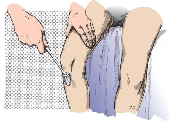
FIGURE 38.8 Method of obtaining the patellar (quadriceps) reflex with the patient seated.
Related posts:

Stay updated, free articles. Join our Telegram channel

Full access? Get Clinical Tree


