Chapter 10 The EEG in Epilepsy
SEIZURE TYPES AND SEIZURE SYNDROME
Seizure Types
The distinction between seizure types and seizure syndromes is central to both the practices of clinical epileptology and clinical electroencephalography. It is worthwhile to consider the distinction between the two and how the diagnosis of each is made. The term seizure type refers to the classification of an individual seizure event. Ideally, a seizure type can be discerned from knowing three key features regarding the event: the patient’s appearance during the seizure, the patient’s subjective description (if any) of what the experience of the seizure was like, and the appearance of the EEG recording made at the time of the event. The details of all three of these features, of course, are not always available to the clinician, especially the simultaneous EEG recording.
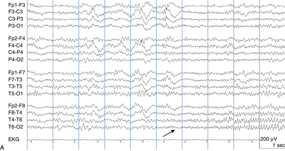
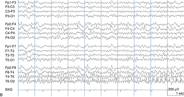
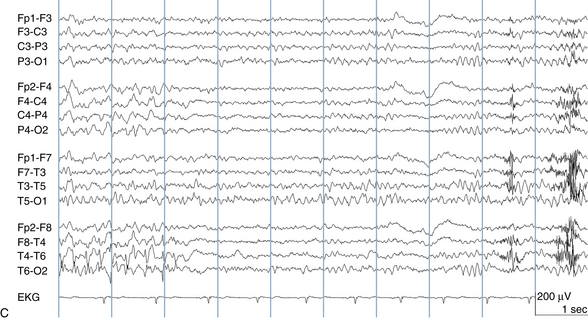
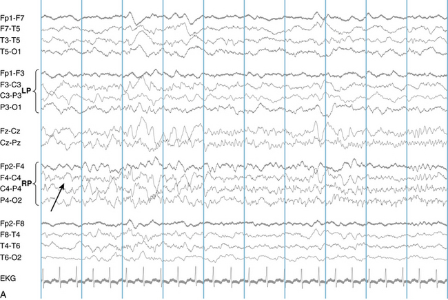
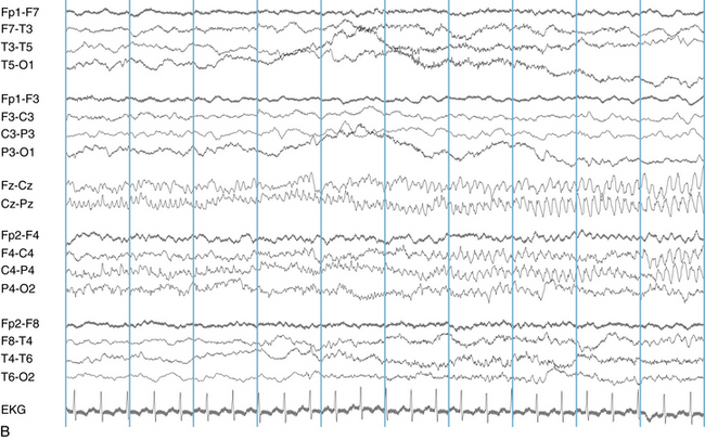
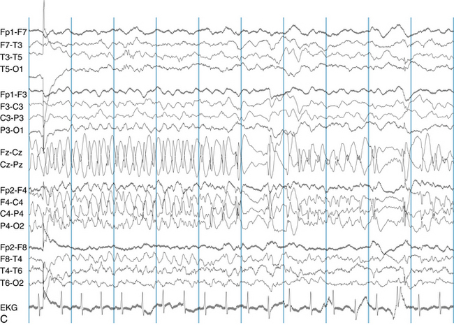
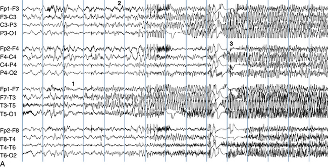
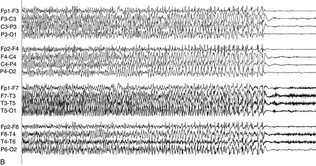
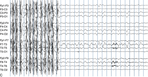
The following hypothetical example illustrates how these three key features are used: a patient experiences an event that starts with a subjective report of a feeling of fear. Next, observers report that the patient begins to stare and is unresponsive, followed by rhythmic jerking of the left arm. Simultaneously, an EEG is recorded that shows a rhythmic discharge starting in the right temporal lobe and evolving to include much of the right hemisphere over a brief period of time (see Figure 10-1, A, B, and C). The combination of psychic aura reported by the patient, staring, the specific motor phenomena reported by observers, and observation of an EEG seizure discharge that starts in the right temporal lobe all establish the diagnosis of a specific seizure type: a complex partial seizure arising from the right temporal lobe. Note that the age and previous history of the patient are not of primary importance in diagnosing seizure type; rather, the behaviors observed during the episode and any recordings made at the time of the event are the most important elements in defining seizure type. We can assign the seizure type “complex partial seizure arising from the right temporal lobe” or “right temporal lobe seizure” without knowing whether the seizures are occurring in a broader context of possible posttraumatic epilepsy or cryptogenic temporal lobe epilepsy. This contrasts with the approach to diagnosing seizure syndromes, described next.


Figure 10-1 A focal seizure beginning in the right temporal lobe is shown (montage setup: left parasagittal chain over right parasagittal chain, left temporal chain over right temporal chain). The arrow indicates the location of seizure onset which consists of a low-voltage, fast rhythm initially confined to the bottom two channels (compare with homologous channels on the left: C3-P3 and P3-O1). This fast rhythm then spreads through the right temporal chain, subsequently involving the right parasagittal chain as well. The discharge slows in frequency and increases in voltage throughout its course before abruptly coming to an end.
Seizure Syndromes
To discern a patient’s seizure syndrome, it is useful to know the patient’s age, history, neurodevelopmental or cognitive status, and the seizure types he or she has experienced. For instance, a 28-year-old man with normal intellect and neurologic examination and a history generalized convulsions and myoclonic jerks since the early teenage years likely has the diagnosis of the seizure syndrome known as juvenile myoclonic epilepsy. In this example, the patient’s seizure syndrome includes two seizure types: myoclonic seizures and generalized tonic-clonic seizures. If necessary, the diagnosis of these seizure types could be confirmed by patient and observer descriptions and simultaneous video/EEG recording of the individual seizure events. The diagnosis of the seizure syndrome of juvenile myoclonic epilepsy, however, is best established by knowing the age at onset of the seizures, the seizure types the patient has, and additional pertinent history.
CLASSIFICATION OF SEIZURE TYPES
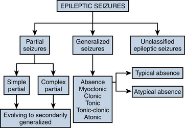
The most frequently used classification of seizure types was established by a committee of the International League Against Epilepsy (ILAE) in 1981. This classification divides seizures into partial and generalized categories and a smaller unclassified category. The process of splitting seizure types up into generalized or partial categories is the first step in seizure classification. Each category of seizure type is described briefly in this section, along with its characteristic clinical and EEG findings. The broad schema of the classification is shown in Figure 10-2.
Partial Seizures
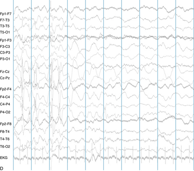
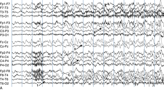
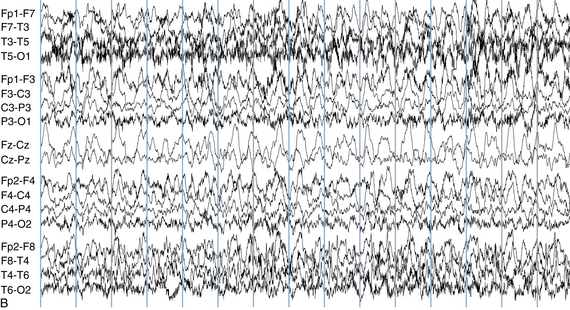
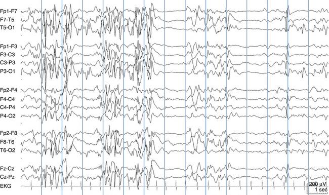
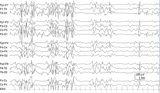
Because a seizure can arise from nearly any location on the cortical surface, the range of potential seizure manifestations is quite diverse. Partial seizures may involve motor findings (such as jerking or stiffening of a limb), sensory findings (such as a sensation of tingling or pain over a region of the body, hearing a sound, smelling an odor, or seeing brightly colored shapes), psychic features (such as a sensation of fear or déjà vu), or autonomic findings (such as sweating or palpitations). It should also be kept in mind that a significant portion of the cortical surface is functionally relatively silent. When a seizure discharge starts in one of these “silent” cortical areas, it is possible for the discharge to occur on the cortical surface without clinical change as a clinically silent seizure rather than a clinical seizure. Frequently, a seizure discharge may begin in one of these “silent” areas and subsequently propagate to other areas such as the motor strip, at which time obvious clinical signs may appear such as clonic jerking of an extremity. Thus, when a patient manifests clonic jerking, we can infer that the seizure discharge has involved the motor strip, but this is not a guarantee that the seizure discharge originated in the motor strip—the seizure discharge may have started elsewhere and propagated to the motor strip. Figures 10-3 and 10-4, show examples of how two focal-onset seizures propagate.


Figure 10-3 This seizure discharge begins in the right parasagittal (RP) area in the form of low-voltage, very fast activity (arrow) and spreads quickly to the midline electrodes (Fz-Cz and Cz-Pz). The seizure onset is detected by noting the first asymmetry between the right parasagittal and left parasagittal (LP) chains. The discharge then becomes most prominent and well formed in the midline channels (Fz-Cz and Cz-Pz) and involves the right parasagittal area, with the field spreading to the right temporal chain before termination.


Figure 10-4 A seizure discharge (arrows) begins in the right temporal chain and right frontal area (montage setup: left parasagittal chain over right parasagittal chain, left temporal chain over right temporal chain). Initially, the seizure waveforms consist of higher voltage sharp forms, but as the seizure spreads to involve the whole of the right hemisphere, it attains a more rounded or sinusoidal morphology. This is an example of a discharge in which the frequency initially accelerates, then decelerates before termination (final termination not shown).
Simple Partial Seizures Versus Complex Partial Seizures
Partial seizures are further subdivided into simple partial seizures and complex partial seizures. A complex partial seizure is a partial seizure associated with diminished consciousness or responsiveness. Some of the partial seizures may be associated with completely retained responsiveness, such as a simple partial seizure involving clonic spasms of the face or hand—the patient may remain completely alert and aware during such a seizure. The majority of partial seizures, however, are associated with diminished responsiveness and are properly classified as complex partial seizures. Indeed, it is only during a minority of partial seizures that complete responsiveness is maintained. During complex partial seizures, the patient may appear awake but is unresponsive (or only minimally responsive) to stimulation. Staring behaviors frequently indicate reduced responsiveness during complex partial seizures. Only rarely does a patient appear to lose consciousness completely during a complex partial seizure.
Epileptic Auras
An epileptic aura is a subjective sensation that heralds the onset of a seizure. The sensations can be diverse, such as hearing a buzzing sound in the ears, experiencing déjà vu, or having a feeling of nausea immediately preceding a seizure. This initial, comparatively minor feature may then be followed by more dramatic seizure manifestations. Strictly speaking, an aura is not a preseizure warning or “prodrome.” Rather, it represents the onset of the seizure itself. The onset of the aura is coincident with the onset of the seizure discharge.
During the aura phase of a seizure, the epileptic seizure discharge is usually confined to a small area, often in the temporal lobe. As it spreads out of its confined area, the clinical seizure manifestations may become more dramatic. Because the aura should properly be considered the onset of a clinical seizure, a true epileptic aura can be counted as a seizure. In some cases, the aura may not progress and represents the seizure in its entirety.
Recordable EEG Patterns Associated With Partial Seizures
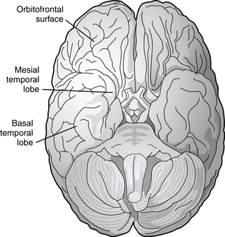
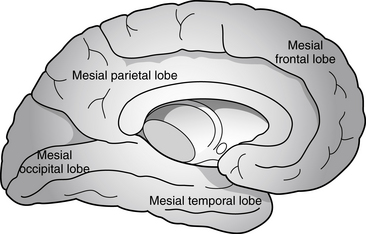
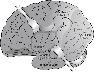
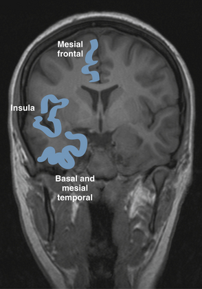
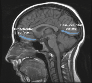
Partial seizures are distinguished by EEG patterns that involve only subsets of the brain. In comparison, the EEG patterns of generalized seizures involve all brain areas at once. Although most epileptic seizures can be recorded using standard scalp electrodes, in some cases a seizure discharge may occur in a cortical area that is not readily accessible to recording with conventional electrodes. These include such areas as the mesial surfaces of the frontal, parietal, and occipital lobes, the orbitofrontal surface of the frontal lobe, the basal occipital and temporal lobes, and the mesial surface of the temporal lobe and insula, among others (see Figures 10-5 through 10-9). Therefore, most, but not all partial seizures are well recorded at the scalp. For these reasons, a negative EEG recording cannot, in and of itself, exclude the diagnosis of an epileptic seizure. In these occasional cases of definite epileptic seizure with negative scalp recordings, other factors must be taken into account to make the correct diagnosis, such as the specific features of the patient event and the history. In cases of partial epileptic seizures associated with a negative EEG, the definition implies that there is some theoretical electrode placement that could record the seizure discharge, even if that placement location would have to be deep within the brain.

Figure 10-5 The basal surfaces of the brain are at some distance from the scalp and difficult to record well using routine techniques. The orbitofrontal surface of the brain lies on the floor of the anterior cranial fossa, above the eyes. The basal temporal lobe lies on the bone of the middle cranial fossa and the mesial temporal lobe, including the amygdala and hippocampus, also lie at some distance from the scalp electrodes.

Figure 10-6 A section through the sagittal midline of the brain shows the mesial surfaces of the frontal, parietal, occipital, and temporal lobes which can be difficult to record.

Figure 10-7 The insula (literally “island”) represents an infolding of cerebral cortex covered by the lips (opercula) of the Sylvian fissure. The frontal and temporal opercula are retracted to reveal this hidden area of cortex.

Figure 10-8 A coronal section of brain is seen on magnetic resonance imaging scan. The mesial frontal lobe, insula, and basal and mesial temporal lobes are highlighted.

Figure 10-9 The basal frontal and occipital lobes are highlighted on a sagittal MRI scan. The gyri of the mesial surface of the cerebral hemisphere (frontal, parietal, and occipital) are also visible.
The EEG patterns associated with partial seizures usually consist of a rhythmic, sharp discharge over the affected area (e.g., spike or spike-wave discharges) as shown in the previous figures. However, partial seizure recordings do not only appear as a train of spikes. Especially when seizure sources are located deeper in the brain and at some distance from the recording electrode, the seizure may only appear as a rhythmic focal slow wave without obvious sharp features when recorded from the scalp. In the case of partial seizures, the epileptic discharge is usually unilateral. Although a unilateral partial seizure discharge may spread incrementally through the primary involved hemisphere, at such time as the discharge might cross to the opposite hemisphere, both hemispheres become simultaneously engulfed with seizure activity; there is no incremental spread through the opposite hemisphere. This process is referred to as secondary generalization.
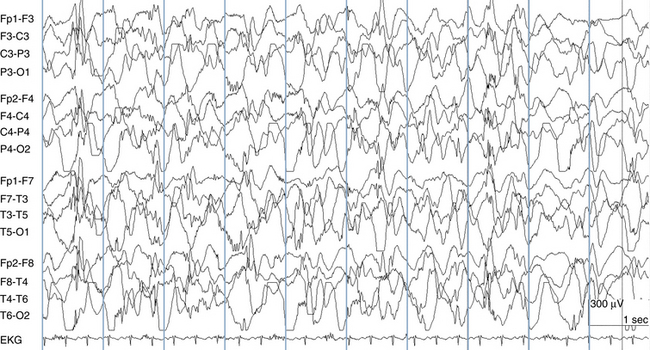
An important exception to this rule is the example of temporal lobe seizures in which the seizure discharge may spread from one temporal lobe to the other without simultaneous involvement of the remainder of the hemispheres (see Figure 10-10). Therefore, in most cases, after the discharge becomes bilateral, the whole of both hemispheres is engulfed with the discharge and the discharge can be considered to have generalized. Complex partial seizures with bilateral temporal involvement represent the exception to this rule: the less typical example of a bilateral seizure discharge that is not truly generalized.


Figure 10-10 The beginning of the low-voltage sharp seizure discharge that is evident on the second half of the first page can be traced back to the right midtemporal area where the muscle artifact stops (1). After 3 seconds, the discharge spreads to the right parasagittal area (2) and then quickly to the midline (3). After 2 more seconds, the discharge has become bilateral and can be seen in the left parasagittal area (4). The sharp waves may be present simultaneously in the left temporal chain as well but would be difficult to discern because of the muscle and motion artifact in that area. As it nears its end, the seizure discharge increases in voltage and slows in frequency. The increased muscle and motion artifact is not unexpected as the clinical manifestation of seizures often includes muscle tensing and patient movement.
Partial Seizures With Secondary Generalization
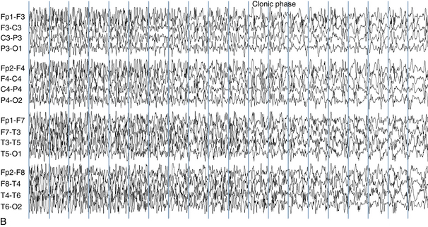
As described earlier, a seizure discharge may start focally and subsequently spread to involve all brain areas (generalize). This flow of the discharge from a subset of cerebral cortex to all of cerebral cortex is reflected both by the spread of the recorded discharge from a subset of EEG channels to all EEG channels and also by an evolution of the patient’s seizure behavior from a partial manifestation to involvement of the whole body. For instance, in the classic example of the type of seizure referred to as a “Jacksonian march,” a patient’s seizure may begin with clonic contractions in the right hand and arm and subsequently spread to the right face and leg. Thereafter, it may spread to the opposite side of the body so that bilaterally synchronous clonic activity of the whole body is seen. This clinical progression is mirrored by an electrographic evolution of the discharge from a small area in the left hemisphere, which includes the portion of the motor strip that is associated with the left hand, to the whole of the left hemisphere and then, finally, to involvement of both hemispheres (see Figure 10-11).


Figures 10-11 A seizure in a child with severe myoclonic epilepsy of infancy (SMEI, or Dravet syndrome) is shown that begins in the left temporal chain (1) and quickly spreads to the left parasagittal chain (2). After a brief period of amplifier blocking (possibly related to patient movement), the discharge becomes bilateral (3). Although SMEI is related to the generalized epilepsies, this patient had similar focal seizure onsets arising from the opposite (right) side at other times.
Partial seizures that secondarily generalize are classified among the partial seizures rather than the generalized seizures for diagnostic reasons. Partial seizures that do not generalize and partial seizures that do secondarily generalize have the same list of possible causes. In comparison, the list of causes of generalized seizures is distinctly different from the list of causes of partial-onset seizures. The tendency to a partial seizure to secondarily generalize usually has little to do with the etiology of the seizure.
Generalized Seizures
The classification of generalized seizures includes absence seizures, myoclonic seizures, clonic seizures, tonic seizures, tonic-clonic seizures, and atonic (astatic) seizures. Even though all of these seizure types fall into the category of generalized seizures, the manifestations of the different types of generalized seizures differ significantly.
Typical Absence Seizures
In its purest form, the sole clinical manifestation of the absence seizure is a pure stare. Absence seizures typically last some 3 to 15 seconds and are characteristically associated with complete unawareness of the environment. In practice, there is no real lower limit to the duration of an absence seizure apart from the ability of the observer or the patient to document unawareness or unresponsiveness for very brief periods of time; it may not be practical to document lack of awareness during discharges that last less than 1 second. Most absence seizures are brief, but they can be of any duration. To distinguish them from atypical absence seizures (described later), the term typical absence seizure may be used. When the “typical” or “atypical” modifier is absent, typical absence seizure is usually assumed. In the past, some have used the term “absence” to denote any seizure associated with staring. In modern usage, the term absence seizure refers to staring seizures associated with generalized spike-wave discharges as described subsequently—complex partial seizures associated with staring are excluded.
A simple typical absence seizure consists of staring alone. The most common modification of the pure stare of the simple typical absence seizure is the addition of rhythmic eye blinking occurring approximately three times per second (each blink occurring in synchrony with the generalized spike-wave discharge seen on EEG). In fact, this sometimes subtle, rhythmic clonic movements of the eyelids occurs with the majority of typical absence seizures; pure staring is relatively uncommon. Less frequent additions to the pure stare of typical absence are clonic or myoclonic movements of the upper body, which also occur in synchrony with the spike-wave discharges, or mild changes in tone during the absence. Although the patient is usually completely unaware of the environment during absence seizures, an occasional patient reports partial awareness and the ability to hear or see during the discharges. In the majority of cases, after an absence seizure, the patient is unaware that the episode has even occurred, the only potential clue being the subjective feeling that something has been missed in the observed sequence of events.
A fraction of patients may have an EEG pattern that is indistinguishable from the 3-Hz generalized spike-wave discharges that occur during clinical absence but may have no change in awareness at all during the discharges. For instance, these individuals may be able to continue a conversation without pause during the discharges, which then, by definition, do not represent clinical seizures (the definition of a clinical seizure requires that an objective or a subjective change occur in the patient at the time of the abnormal EEG discharge). Realizing that this phenomenon exists, the electroencephalographer must resist the impulse to assume that all 3-Hz generalized spike-wave discharges represent absence seizures. Even “classic” 3-Hz generalized spike-wave discharges may occur as an interictal abnormality.
Automatisms
Especially during lengthier absence seizures, individuals can manifest automatic behaviors during the seizure discharge. The expression of such automatisms (which may also be seen during complex partial seizures) varies widely. Examples include fumbling of the hands, running the fingers through the hair, or making humming sounds. Unlike other clinical features of the seizure, such automatisms are not driven directly by a seizure discharge in the way that clonic jerking would be driven by repetitive spikes in the EEG. Rather, automatisms represent the release of automatic, preprogrammed behaviors. When the cerebral cortex is functioning normally, these automatic programs are suppressed. When the cerebral cortex is involved with seizure activity, however, as during an absence seizure, such preprogrammed behaviors may be released.
EEG
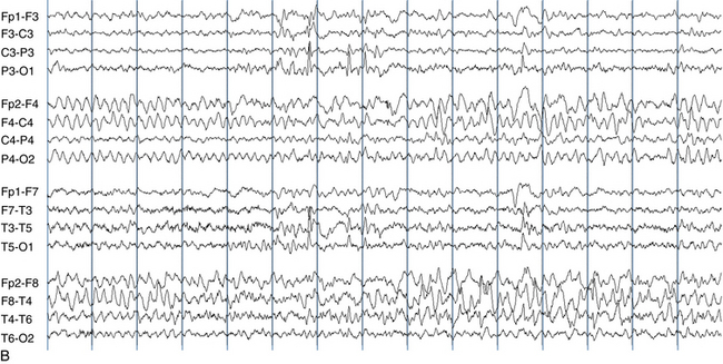
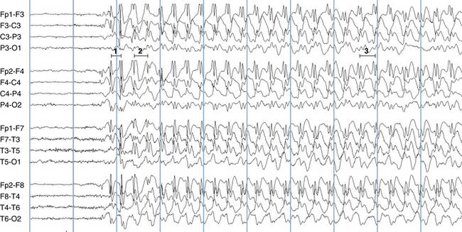
The most frequent EEG correlate to typical absence seizures is the “classic” 3-Hz generalized spike-wave discharge (see Figure 10-12). When these discharges are analyzed closely, the maximum voltage of the spike component of the spike-wave complexes is most commonly seen in the superior frontal electrodes (F3 and F4). Less often, the spike maximum is seen in the occipital area, and even less frequently in other locations.

Figure 10-12 This 3-Hz generalized spike-wave discharge shows the abrupt onset and termination that is characteristic of absence seizures. Note the frontal maximum of the waveforms.
Although the term 3-Hz generalized spike wave is well known and implies a consistent frequency, observed firing frequencies are not necessarily as consistent as the term implies. Often, the first few discharges fire at a frequency slightly faster than 3 Hz. After onset of the discharge, the firing frequency typically slows, often to 2.5 Hz and sometimes to 2 Hz before abruptly terminating (see Figure 10-13). One of the most characteristic attributes of the typical absence seizure is the abrupt onset and termination of the discharge. The classic 3-Hz generalized spike-wave discharge tends to occur against a normal background and has a clear time of onset and a fairly well demarcated termination. After termination, the EEG returns to the previous background after a few seconds or less.

Figure 10-13 Although the discharge associated with this typical absence seizure is classified as 3-Hz generalized spike-wave, note that the discharge’s firing frequency still evolves throughout its course. The first wavelength measured suggests a firing rate just above 4 Hz but (1), 1 second later at the time of the second measured wavelength, the firing rate has dropped to 3 Hz (2). Later in the discharge, the third measured wavelength implies a firing frequency of 2.5 Hz (3).
Atypical Absence Seizures
As with typical absence seizures, the main clinical features of atypical absence seizures are staring and unresponsiveness. Atypical absence seizures differ, however, in that onset and termination of the episodes, both clinically and electrographically, are less clear, and firing rates are slower. Also, atypical absence seizures tend to occur in individuals with cognitive impairment or mental retardation, whereas typical absence seizures are more often seen in subjects who are cognitively normal. Changes in tone are more common during atypical absence seizures, with slumping of the head, shoulders, and sometimes the whole torso seen during some examples.
EEG
The EEG hallmark of atypical absence seizures is the slow spike-wave discharge. Slow spike-wave discharges differ from “classic” 3-Hz generalized spike-wave discharges in two important respects: slow spike-wave discharges, as their name implies, fire at a slower rate, usually 2.5 Hz or less at onset; see Figure 10-14). Slow spike-wave discharges also lack the clear-cut onset and termination characteristic of typical absence seizure discharges. Finally, whereas slow spike-wave discharges are often generalized, asymmetries, both between the left and the right hemispheres and the anterior and posterior head regions, are more common. There is no single characteristic location for the discharge maximum for the slow spike-wave discharges associated with atypical absence seizures, and the location of the voltage maximum may differ even within the same patient at different times. Atypical absence seizures often occur against the backdrop of an otherwise abnormal EEG, which may include scattered epileptiform activity or a slowed background.

Figure 10-14 Slow spike-wave discharges are seen in a young man with mixed seizures. Note the slowed firing rate of the train of spike-wave discharges on the second half of the page and the scattered, single discharges in multiple locations seen on the first half of the page.
Slow-spike wave discharges are often associated with atypical absence seizures, but this is not always the case. Although slow spike-wave discharges are expected as the EEG correlate of atypical absence seizures, the converse is often not true: most slow spike-wave discharges are not associated with clinical atypical absence seizures. In practice, slow spike-wave discharges are often seen as interictal abnormalities in the EEG. The simple observation of slow spike-wave discharges in the EEG is no guarantee that the patient is actually experiencing an atypical absence seizure, although the finding does raise suspicion that the patient has this seizure type. When this pattern is seen, the concurrent observation of associated staring or some form of decreased responsiveness or change in tone is necessary to establish the diagnosis of an electroclinical seizure. This same phenomenon of electrical discharge without clinical change may also occur with “classic” 3-Hz generalized spike-wave discharges as described in the previous section, although much less often.
Myoclonic Seizures
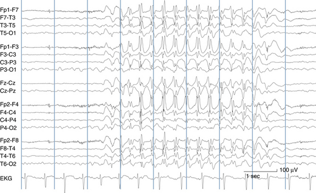
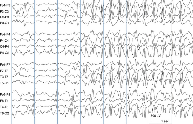
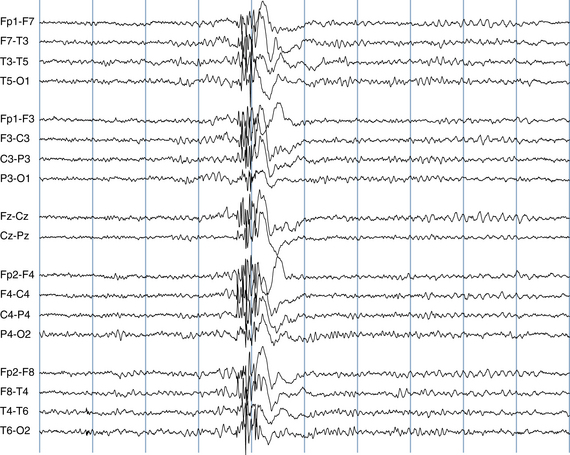
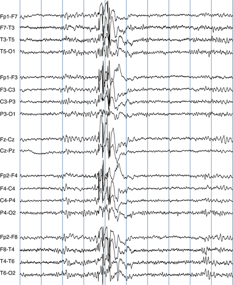
Myoclonic seizures consist of a lightning-like or shock-like contraction of the muscles driven by an epileptic discharge. The appearance of a myoclonic seizure is similar to experiencing a brief electric shock. Myoclonus may consist of a single jerk or a quick series of jerks that occur in a burst, either rhythmic or nonrhythmic. Epileptic myoclonus may manifest as a muscle jerk in nearly any part of the body, although the most common location for epileptic myoclonus is the upper shoulder girdle. In such cases, the myoclonus usually consists of a quick series of abduction jerks at the shoulders. During the series of jerks, there may be a tendency for slight net abduction of the upper arms away from the body with each jerk. The most common EEG manifestation of epileptic myoclonus is a high-voltage polyspike-wave discharge, which may occur singly or in brief, repetitive bursts (see Figures 10-15 and 10-16).

Figure 10-15 This 15-year-old girl was referred for tremor. The movements in question actually represented epileptic myoclonus driven by the high-voltage polyspike-wave discharge shown.

Figure 10-16 Myoclonic jerks often occur in quick succession. In this teenage patient, this quickly repetitive series of polyspike-wave discharges caused a series of quick abduction jerks at the shoulders.
It is important to keep in mind that not all myoclonus is epileptic. Nonepileptic myoclonus may originate in the central nervous system at levels below cerebral cortex, including the subcortical areas, brainstem, and even the spinal cord (segmental myoclonus). The question of whether an instance of myoclonus is epileptic myoclonus is best confirmed by demonstrating the presence of a concomitant EEG discharge driving the movement. No EEG discharge would be expected to accompany nonepileptic myoclonus.
Clonic Seizures, Tonic Seizures, and Generalized Tonic-Clonic Seizures
Clonic Seizures
Clonic seizures are characterized by repetitive clonic jerks. These clonic jerks can occur in nearly any skeletal muscle group in the body depending on the cortical location of the discharge (usually including the motor strip). Clonic jerking from seizure activity tends to have a rhythmic quality. Because each clonic jerk is driven by an EEG discharge, a simultaneous spike or spike-wave discharge is expected with each clonic jerk. Therefore, the frequency of the EEG discharges typically matches the frequency of the jerks. Because clonic jerking is typically driven by discharges from the area of the motor strip, it is rare that scalp EEG electrodes will fail to record them, especially if they involve the face or hand.
Tonic Seizures
Tonic seizures cause tonic stiffening of a limb, several limbs, or the whole body. When the whole body is involved with a generalized tonic seizure, tonic stiffening in extension is most common; however, tonic stiffening with flexion of the hips, knees, or arms may also be seen. Especially with generalized tonic seizures, tonic contraction of the diaphragm may occur resulting in a forced inhalation or grunting sounds. Most often the EEG correlate of tonic seizures is a spray of rapid spikes in the affected area, often with an initial frequency of 10 to 25 Hz, which subsequently slows, similar to the onset of tonic-clonic seizures as described next. Other EEG correlates are not uncommon, including an abrupt desynchronization (flattening) of the EEG. When such flattening is seen, there is sometimes a suggestion of low voltage rapid spikes superimposed on the flattened pattern though these are not always identifiable (see Figure 10-17). Therefore, although most ictal patterns are dramatic and show high-voltage repetitive discharges, the electroencephalographer must also be alert to abrupt flattening of the EEG (an electrodecrement) as a seizure correlate. Other patterns are less common.
Generalized Tonic-Clonic Seizures
The generalized tonic-clonic seizure refers specifically to the sequence of whole-body tonic stiffening followed by clonic jerking. Unfortunately, this term is often used indiscriminately to refer to any generalized convulsion, a use which is technically incorrect; properly, the term generalized tonic-clonic seizure should be reserved exclusively for the sequence of whole-body stiffening followed by whole body clonic jerking. This distinction can be important as some clinical events which mimic seizures, such as convulsive syncope (nonepileptic stiffening and jerking body movements associated with brief episodes of significant hypotension, do not tend to manifest this specific sequence. The sequence of tonic body stiffening followed by clonic jerking, compared with other possible sequences of movements, is highly suggestive of epileptic seizure and should be duly noted.
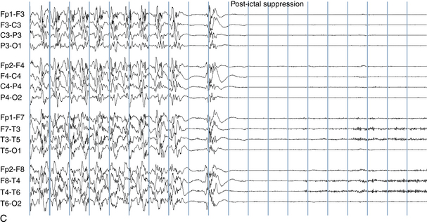
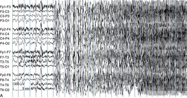
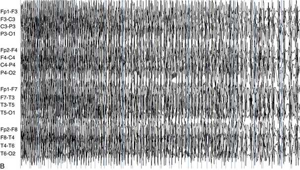
The EEG correlate of the tonic-clonic seizure often begins with an abrupt onset of generalized rapid spikes which then slow in frequency over the course of the event. As the firing frequency of the spikes slows, the spikes may begin to manifest a clearer spike-wave morphology. From the clinical perspective, rapid spikes are often associated with tonic stiffening. At a certain point in the seizure, the firing rate of the spikes slows to a point that allows each spike or spike-wave discharge to generate a separate clonic jerk (see Figure 10-18). This is the reason that the progression from tonic stiffening to clonic jerking is so common. Over the course of a generalized tonic-clonic seizure the clonic jerking is seen to slow in frequency and blend into a slow-wave pattern: “postictal slowing.” Less commonly, a clonic-tonic-clonic seizure may occur in which clonic jerking speeds up and melds into tonic stiffening, followed by the usual progression back to clonic jerking. As expected, the EEG correlate of this type of seizure often consists of spike-wave discharges that speed up to become rapid spikes, and then slow down again (see Figure 10-19).


Figure 10-18 The classic evolution of a generalized tonic-clonic seizure is shown. There is abrupt onset of generalized rapid spikes at the start of the tonic phase. As the firing frequency of the spikes decreases the individual spikes become far enough apart from one another that each spike can generate a separate clonic jerk, representing the clonic phase of the seizure. In this case, there is a period of postictal suppression after cessation of the seizure discharge. After seconds or minutes, generalized slow-wave activity appears that may last from minutes to days depending on the duration of the seizure and the nature of the patient.


Figure 10-19 A clonic-tonic-clonic seizure discharge is shown. Note that, in comparison to the previous example, the initial rapid spikes appear in brief bursts (seconds 8–10 of panel A) each associated with a clonic jerk. After a few seconds, the rapid spikes consolidate and their firing frequency increases, corresponding to the tonic phase of the seizure. The seizure discharge then follows a pattern similar to that of the tonic-clonic seizure shown earlier, with slowing of the spike frequency associated with the clonic phase of the seizure. Postictal slowing is seen following this seizure discharge.
Atonic Seizures
As the name implies, atonic seizures consist of a loss of tone, usually of the truncal muscles. In its mild form, an atonic seizure may simply consist of a subtle slumping of the shoulders. More frequently, an atonic seizure may manifest as a head-drop spell in which the patient’s head slumps forward. The most dramatic version of an atonic seizure is the drop attack in which the patient collapses to the ground, possibly resulting in injury. An astatic seizure is a seizure resulting in a fall (discussed further in the section on seizure types in Lennox-Gastaut Syndrome). There are many possible EEG correlates to atonic seizures, including slow spike-wave discharges, EEG desynchronization (flattening), or polyspikes, sometimes followed by flattening.
Unclassified Seizure Types
There a few seizure types that do not fit into the foregoing classification. These include swimming or bicycling movements, apneas, and roving eye movements. In fact, it remains controversial as to whether many of these apparent seizure types represent true epileptic phenomena. It is possible that some of these behaviors represent automatic movements (automatisms) rather than seizure activity driven by an actual epileptic seizure discharge. The nature of such events therefore remains to be clarified. Infantile spasms (or epileptic spasms) represent a seizure type that is not easily classified as generalized or focal (see the later section on West syndrome).
Neonatal Seizures
Neonatal seizures have been given a classification scheme different from the general ILAE seizure classification described earlier. The most common neonatal seizure classification system in use today was described by Volpe in 1989. The seizure types described in this classification are actually quite similar to those of the ILAE classification, with the exception of an additional seizure category referred to as “subtle seizures,” a subgroup that remains controversial as the epileptic nature of subtle seizure behaviors has not yet been firmly established. See Chapter 13, “The EEG of the Newborn” for further discussion.
CLASSIFICATION OF SEIZURE SYNDROMES
A patient’s seizure syndrome is defined by the type or types of seizures experienced, age of onset, neurologic status (abnormal neurologic status before or after seizure onset), progression, family history, physical examination, and EEG patterns. Identification of a particular seizure syndrome will often suggest possible treatments and a specific prognosis. Select seizure syndromes are discussed below, generally in order of age of onset.
Epileptic Syndromes of Early Infancy Associated With a Burst-Suppression Pattern
Early myoclonic epilepsy (EME) and early infantile epileptic encephalopathy (EIEE) are the two major catastrophic epilepsies of early infancy. Although these two syndromes have much in common, they appear to represent two distinct entities.
Early Myoclonic Encephalopathy
Infants with EME present soon after birth with both fragmentary and massive myoclonic seizures. Other seizure types also occur. The magnetic resonance imaging (MRI) scan at birth is almost always normal, and a large fraction of these babies are eventually found to have a specific metabolic disorder, nonketotic hyperglycinemia (NKH). Those babies with EME who do not prove to have NKH may have other metabolic diagnoses, and it is felt that the remaining cases of EME may be caused by some metabolic entities yet to be defined. This belief is based on the observations that EME babies have anatomically normal brains by MRI, and there is usually no history of a previous neurological injury. Babies with EME have complete developmental failure, and the seizures tend to be refractory to treatment. The EEG shows an unremitting burst-suppression pattern that may continue unabated through childhood (see Figure 10-20).

Figure 10-20 A dramatic burst-suppression pattern is seen in the EEG of this newborn with early myoclonic epilepsy (EME). No sleep cycling occurred in this recording, and every page of the record showed the same burst-suppression pattern. In patients with EME, this discontinuous pattern may persist for years.
Early Infantile Epileptic Encephalopathy
EIEE, also known as Ohtahara syndrome, appears to be a “lesional” epilepsy syndrome and, in contrast to EME, is often associated with an abnormal MRI scan. Tonic seizures are more prominent in EIEE compared with EME. MRI abnormalities associated with this syndrome can include cerebral malformations, or cerebral injuries as may occur in babies with hypoxic-ischemic encephalopathy. Therefore, EIEE occurs as an epileptic syndrome that is believed to be symptomatic of a preexisting abnormality, be it a cerebral malformation or some type of brain injury. EIEE is more likely to evolve to West syndrome or the Lennox-Gastaut syndrome.
Like EME, EIEE is typically associated with a burst-suppression pattern on EEG. The EEG patterns of EIEE and EME are not easily distinguished without the benefit of the clinical history (see Figure 10-21). The burst-suppression pattern of EIEE is more likely to evolve into other EEG background patterns later in life, compared with the EME burst-suppression pattern, which may persist indefinitely.

Figure 10-21 The burst-suppression pattern of a newborn patient with EIEE shown in this figure is essentially indistinguishable from the patterns seen in EME. In EIEE, eventual evolution to other patterns over months or years is not uncommon.
Therefore, despite the many aspects they share in common (similar EEG pattern, intractable seizures, poor prognosis), EIEE distinguishes itself from EME in that EIEE is considered an acquired or lesional epileptic encephalopathy caused by a cerebral malformation or a cerebral injury. In contrast, children with EME are believed to have the disorder on a genetic or biochemical basis rather than from a postnatal event. In EME, MRI brain anatomy is typically normal.
The Concept of the Age-Dependent Epileptic Encephalopathies
EIEE is considered one of the age-dependent epileptic encephalopathies. This group of syndromes represents the result of different characteristic epileptic responses of the brain to injury seen at different ages. For instance, when the brain is injured in the neonatal period, the response to injury may present as the EIEE syndrome as described earlier. When the epileptic response to an injury or abnormality occurs later in infancy (typically after 3 months of age), the child may develop a pattern of infantile spasms or West syndrome (discussed later). The epileptic response to an injury that appears after 3 years of age may present as the Lennox-Gastaut syndrome (discussed later). Because different maturational states of the brain are only associated with certain syndromic patterns, West syndrome does not present in adults, and Lennox-Gastaut syndrome cannot present in early infancy; because of the way the human brain matures, it is not able to mount those types of seizure patterns at those ages.
Pyridoxine-Dependent Seizures
The syndrome of pyridoxine-dependent seizures is extremely rare in its pure form. In the classic presentation of this disorder, a newborn is found to have refractory seizures associated with a persistent burst-suppression pattern on EEG. Administration of pyridoxine, sometimes in high doses, abruptly breaks the abnormal EEG pattern and promptly terminates the seizure activity. Without continued supplementation with pyridoxine, the seizures will relapse. Because this is such an easily treatable form of what would otherwise be a catastrophic epilepsy, pyridoxine infusion is routinely carried out on newborns with seizures who potentially match the phenotype of this disorder (in particular, those having a burst-suppression pattern on EEG without other explanation). Later onset and partial forms of this disorder exist and are even less commonly encountered.
Benign Familial Neonatal Convulsions and Benign Neonatal Convulsions
Benign familial neonatal convulsions (BFNC) and benign neonatal convulsions are the two important benign seizure syndromes of the newborn. The essence of both of these seizure syndromes is similar: seizures early in the newborn period in a previously well infant followed by a generally benign outcome. Although these two syndromes have significant similarities, they are discussed separately.
Benign Neonatal Convulsions
The syndrome of benign neonatal convulsions is also known to as “fifth day fits,” a name that serves as a useful reminder that the fifth day of life is the most common age of onset for this syndrome. To some extent, the diagnosis of benign neonatal convulsions must be made in retrospect because a benign long-term course is a key part of the syndrome.
The expected presentation of benign neonatal convulsions consists of a newborn who appears normal at birth and who may have already been uneventfully discharged home from the hospital. The seizures begin in the first week of life, with the most common age of incidence being the fifth day of life; 90% of babies present between the fourth and sixth days of life. The seizures generally subside by the second month followed by continued normal development.
The family history for seizures is negative. There is no antecedent history of a difficult delivery or birth injury, and neuroimaging is normal. Apart from possible mild hypotonia, the interictal examination is normal. A search for central nervous system infection is negative, and no electrolyte or other metabolic disturbances are found. The clinician is left with a story of an otherwise perfectly well newborn with unexplained onset of seizures in whom all testing is normal.
In babies with benign neonatal convulsions, the EEG background pattern tends to be normal. A characteristic EEG finding has been described in such babies, termed théta pointu alternant. This is a pattern of sharpened theta waves occurring in brief runs, typically in each central area, and alternating sides (see Figure 10-22). Although this pattern is said to occur in the majority of patients with this seizure syndrome, it may be difficult to identify, and its presence is not necessarily diagnostic of benign neonatal convulsions.

Figure 10-22 Théta pointu alternant, or alternating sharp theta, may be seen in each central area in both the syndromes of benign neonatal convulsions and benign familial neonatal convulsions.
(Modified from Dehan M, Quillerou D, Navelet Y, et al. Convulsions in the fifth day of life: a new syndrome? Arch Fr Pediatr 1977;34:730 742).
Benign Familial Neonatal Convulsions
The syndrome of benign familial neonatal convulsions (BFNC) has many elements in common with the previously described syndrome of benign neonatal convulsions. As the name implies, however, in such babies there is a positive family history of seizures in the newborn period. In this syndrome, the seizures tend to begin slightly earlier, typically on the second or third day of life, usually resolving by the second month. Unlike benign neonatal convulsions, in BFNC a 10% to 15% incidence of later epilepsy has been described. Also, mild developmental problems may occur with a slightly increased frequency compared with the unaffected population. A large proportion of affected individuals have been found to have linkage to the 20q13.3 gene locus corresponding to a mutation in the potassium channel gene KCNQ2. A smaller number of affected individuals have been found to have linkage to the 8q24 locus and a mutation in the KCNQ3 gene (both potassium channel genes). Still other kindreds appear to have no abnormality at either of these loci, suggesting that additional genetic abnormalities that cause this syndrome have yet to be identified.
The typical seizure in BFNC is the clonic seizure, preceded by tonic stiffening and apnea in some (Hirsch et al., 1993). Most commonly the interictal EEG is normal, however, the théta pointu alternant pattern, as has been described in benign neonatal convulsions, has been reported in some babies with this syndrome.
Febrile Seizures
Febrile seizures are seizures that occur with fever during childhood. There are mild variations in the age range that various groups have used to define febrile seizures, with ranges such as 3 months to 5 years and 1 month to 6 years being used. These stated age ranges are somewhat misleading in that it is uncommon for febrile seizures to start at the end of these age ranges; usually when a febrile seizure occurs after the age of 4 years, the child has already had previous episodes. The large majority of affected children have had the first febrile seizure by 3 years of age. According to the definition, the seizure should not have an obvious cause such as central nervous system infection, and children with previous unprovoked seizures are excluded from the definition. The fever should exceed 38.4 °C (101 °F), but this cutoff is flexible. The diagnosis of febrile seizures is usually made in a normal-appearing child with a normal nervous system. Nevertheless, there is no reason that children with preexisting neurologic abnormalities should be any less prone to febrile seizures than their normal counterparts leaving the underlying cause of the seizures more difficult to sort out in this group.
Although one may speak of a “febrile seizure syndrome,” febrile seizures are generally not considered an example of an epilepsy syndrome. Epilepsy is defined as a tendency to recurrent, unprovoked seizures, and because the presence of fever is considered a provocative factor, febrile seizures are not considered examples of epileptic (unprovoked) seizures.
Febrile seizures are common. They occur in 3% to 5% of all children and, in the large majority, disappear during childhood. Only 2% to 4% of children with febrile seizures are destined to have future seizures without fever (epilepsy). Fever is known to be a common seizure-triggering factor for persons who do have epilepsy, and the question may arise as to whether an apparent febrile seizure episode really represents an epileptic seizure triggered by fever in a child who is destined to have epilepsy. It is known that certain features of a febrile seizure episode increase the odds that a child will later develop epilepsy. A febrile seizure is termed a complex febrile seizure when one or more “complex” features are present. These complex features have been defined by epidemiologic studies that have found these factors to be associated with an increased risk of developing later epilepsy (afebrile seizures). They include seizure duration longer than 10 or 15 minutes, focal (as opposed to generalized) febrile seizures, or two or more seizure episodes within a 24-hour period. The more of these “complex” features a child has, the higher the risk of developing later epilepsy. A preexisting abnormal neurological status at the time of seizure onset and a positive family history of epilepsy also independently increase the chance of later epilepsy in a child with febrile seizures.
A relationship between febrile seizures and temporal lobe epilepsy has long been suspected. Case-control studies of individuals with temporal lobe epilepsy appear to show an increased incidence of a history of febrile seizures, particularly prolonged febrile seizures, in temporal lobe epilepsy patients compared with control subjects. These findings suggest the possibility that prolonged febrile seizures may cause hippocampal damage (hippocampal sclerosis) and predispose to later temporal lobe epilepsy. An alternative interpretation is that those children destined to have temporal lobe epilepsy later in life may automatically be more prone to prolonged febrile seizures in childhood and that the early febrile seizures may not be a causative factor in the later epilepsy.
The Role of EEG in the Evaluation of Febrile Seizures
Despite the fact that EEG abnormalities may be seen in children with febrile seizures, the EEG has not proved to be a particularly useful tool in predicting later epilepsy in these children. The most common epileptiform abnormality found in children with febrile seizures is generalized spike-wave discharges. This pattern is, of course, nonspecific in that it is also seen in the generalized epilepsies. Its presence in the EEG of a child with febrile seizures has not been clearly shown to increase the risk of later epilepsy. Increased slowing, especially in the posterior quadrants, may be expected within a few days of a febrile seizure. In general, because the EEG has not been found to be useful in predicting later epilepsy in children with febrile seizures, EEG testing is not routinely indicated in the evaluation of uncomplicated febrile seizures. Also, because febrile seizures are rarely treated with daily antiepileptic medications, it is highly unlikely that an EEG result, whether or not it is abnormal, would prompt the use of daily seizure medication.
Infantile Spasms and West Syndrome
Although the terms infantile spasms and West syndrome are sometimes used interchangeably, infantile spasms specifically refers to a seizure type (most often, but not exclusively seen as a part of West syndrome) and West syndrome to an epilepsy syndrome. The term West syndrome denotes a syndrome consisting of the triad of infantile spasms, an EEG pattern of hypsarrhythmia, and neurodevelopmental abnormality in childhood. The original clinical description of this syndrome appears in a letter to Lancet by Dr. W. J. West in 1841, who described the seizures in his son.
The hallmark seizures of West syndrome are infantile spasms. Infantile spasms may occur as flexor spasms, extensor spasms, or mixed (asymmetrical) spasms. Flexor spasms are the most common form, consisting of a brief tonic contraction in flexion of the body on the hips, flexion of the head on the neck, and tensing of the shoulders, sometimes in abduction. Because of the flexed position attained during the seizures, these episodes have also been referred to as “jackknife seizures” or “Salaam seizures.” The position is typically held for approximately 1 second, followed by relaxation. The episodes tend to occur in clusters; a series of repeat spasms may last several minutes. Less commonly, spasms can result in extensor rather than flexor posturing. The presence of asymmetrical spasms should always prompt a search for an underlying focal lesion. Other, milder variants of spasms can be seen, including relatively subtle bobbing of the head with upward eye deviation and mild shoulder movement. Because epileptic spasms are seen in both apparently generalized and focal forms, this seizure type defies easy categorization into one group or the other.
Infantile spasms are occasionally erroneously classified as myoclonic seizures, but the episodes are not consistent with the lightning-like jolt that is the definition of myoclonus. Rather, the clinical spasm usually lasts approximately 1 second, too long to be classified as myoclonus. Although the large majority of spasms occur during infancy, occasionally spasms persist past infancy. In older children or adults, the term infantile spasms becomes awkward, and the term epileptic spasms is preferred.
The infantile spasms of West syndrome can be surprisingly difficult to diagnose. Because West syndrome is a rare disorder, many pediatricians have not encountered this seizure type in clinical practice. The tensing up seen during infantile spasms can mimic gastrointestinal discomfort or episodes of colic. Often, the primary care physician does not have the opportunity to witness the seizures personally but must rely on a verbal description of flexing up of the knees followed by crying, a history that may not initially suggest seizures. Features of the history that increase the suspicion of infantile spasms include the short duration of the individual tensing movements, the tendency for the episodes to cluster, and the tendency for the clusters to occur in the period after awakening from sleep.
The infantile spasms of West Syndrome usually begin between the ages of 3 and 18 months. Only rarely is onset of this seizure type seen outside of childhood. In many, the seizures resolve spontaneously but are often later replaced by other seizure types, such as focal seizures or the mixed seizures of the Lennox-Gastaut syndrome. Only a relatively small minority of patients are intellectually normal after developing infantile spasms and hypsarrhythmia.
EEG in the Diagnosis of Infantile Spasms and West Syndrome
Because West syndrome has distinctive ictal and interictal EEG signatures, electroencephalography is a central tool in the diagnosis of infantile spasms and West syndrome. EEG helps establish the diagnosis of infantile spasms, either by identifying the characteristic interictal pattern of West syndrome (i.e., hypsarrhythmia) or by recording the spasms and demonstrating an ictal discharge during the events. EEG may also be used to monitor the success of treatment.
The EEG term hypsarrhythmia is derived from the Greek meaning “high” or “lofty” rhythm. In fact, some of the highest voltages measured in electroencephalography are seen in babies with hypsarrhythmia. Compared with adult EEGs in which voltages typically do not exceed 200 μV, hypsarrhythmia EEGs may exceed 1 mV (1000 μV). The essential features of the hypsarrhythmic pattern are high voltage and chaos. In this context, chaos refers to the opposite of synchrony. A completely chaotic EEG pattern is a pattern in which different electrical events and rhythms are occurring in different brain regions at different times in an unsynchronized and seemingly unrelated fashion. In contrast, the generalized spike-wave discharge, although abnormal, represents a pattern with a high degree of synchrony with all cortical areas acting in unison. In the chaotic hypsarrhythmia pattern, rapid spikes, high-voltage s low waves, and other abnormalities may occur in scattered locations at different times and in a seemingly random fashion (see Figure 10-23). Intermediate states between synchrony and complete chaos are also seen (see Figure 10-24).

Figure 10-23 The high-voltage, chaotic pattern of hypsarrhythmia is shown. Note the “random delta” activity and the scattered spikes, occurring both singly and in brief runs in a chaotic fashion.

Stay updated, free articles. Join our Telegram channel

Full access? Get Clinical Tree

