The Myoclonic Epilepsies


The Myoclonic Epilepsies


Renzo Guerrini

Paolo Bonanni

Carla Marini

Lucio Parmeggiani


The term myoclonic has traditionally designated a large group of epilepsies characterized by repeated brief jerks, often responsible for multiple falls, by severe seizures resistant to antiepileptic drugs, and by an association with cognitive impairment (1,2). However, not all the ictal manifestations that cause falls are myoclonic, and not all myoclonic epilepsies predict a poor outcome. Myoclonic jerks are the only seizure type in but a minority of patients with myoclonic epilepsy (1,2), which is commonly associated with generalized tonic-clonic seizures (2,3), as well as generalized clonic, atypical absence, and atonic seizures (4,5). Tonic seizures are uncommon in myoclonic epilepsies, but isolated tonic attacks during sleep are not rare in children with myoclonic-astatic epilepsy (MAE) (1,6). As a result, confusion arose in classifying myoclonic epilepsies, because they represent a broad group of diseases and epilepsy syndromes that differ in evolution and prognosis. This chapter defines clinical and electroencephalographic (EEG) features of myoclonic seizures, distinguishes them from other seizure types, and describes diagnosis and treatment of various syndromes with prominent myoclonic seizures, including progressive disorders.


DEFINITION

Myoclonus, which is used to describe involuntary, jerky movements frequently involving antagonist muscles (7), can be classified physiologically as epileptic and nonepileptic. Epileptic myoclonus is an elementary electroclinical manifestation of epilepsy involving descending neurons, whose spatial (spread) or temporal (self-sustained repetition) amplification can trigger overt epileptic activity (1). Myoclonus can have a focal, multifocal, or generalized distribution (8). Epileptic myoclonus is characterized neurophysiologically by a myoclonic electromyographic (EMG) burst ranging between 10 and 100 milliseconds, synchronous EMG bursts or silent periods on antagonist muscles, and an EEG correlate detectable by routine surface electroencephalography or burst-locked EEG averaging (1).


CLINICAL AND ELECTROENCEPHALOGRAPHIC FEATURES

Myoclonic seizures can produce only slight head nodding or abduction of the arms (or both) or be responsible for falls when the lower limbs are also involved or when the atonic phenomenon immediately after the jerk is prominent (3,9). Myoclonic seizures are short and usually cluster, with a repetition rate of 2 to 3 Hz, and most commonly affect axial muscles, but in 57% of patients, may also involve the extraocular muscles, eyelids, and face, especially perioral muscles (10). Most occur spontaneously on awakening, as in juvenile myoclonic epilepsy (JME), or during drowsiness, as in benign myoclonic epilepsy. Jerks may sometimes be precipitated by photic stimulation in the electroencephalography laboratory or induced by natural photic stimuli. Tapping or sudden acoustic stimuli may also cause generalized myoclonic jerks of epileptic origin (11). Usually isolated events, myoclonic seizures at times are repeated in prolonged series and, in some cases, can progress to a myoclonic status with partial preservation of consciousness (5,12).

The electromyographic (EMG) recording shows a biphasic or polyphasic potential lasting from 20 to 150 milliseconds. Generalized bursts of polyspike and waves are evident on ictal electroencephalography. The interictal recording may be entirely normal or show slow background activity, depending on whether the myoclonic attacks are part of an idiopathic or a symptomatic epilepsy. Focal abnormalities are rare. Interictal bursts of irregular polyspike waves lasting
less than 3 seconds can be spontaneous or induced by photic stimulation (2,5). Discharges featuring multiple generalized spikes and waves often increase in frequency during nonrapid eye movement (NREM) sleep (2,5).


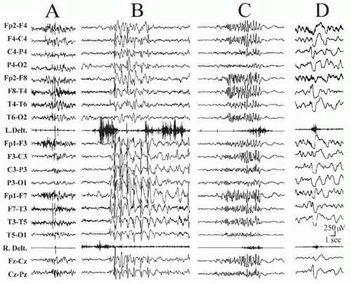
Figure 26.1 Types of brief seizures that clinically manifest as body jerks, shown by their neurophysiologic correlates. A: Myoclonic seizure; electromyographic (EMG) burst lasts less than 100 milliseconds and is time-locked to a polyspike-and-wave discharge. B: Atonic seizure; a spike-and-wave discharge is accompanied by an EMG silent period lasting as long as the discharge on scalp electroencephalography. Note the progressive onset of the silent period and the transient resumption of muscle tone, resembling a myoclonic jerk, interrupting the EMG silence. C: Brief tonic seizure; a polyspike discharge is associated with a bilateral tonic contraction of both deltoid muscles lasting about 5 seconds, that begins with a crescendo pattern. D: Epileptic spasm; a brief (approximately 500 milliseconds), diamondshaped EMG burst correlated with a high-amplitude, diffuse, polyphasic slow transient on EEG channels.


DIFFERENTIAL DIAGNOSIS

The recognition and classification of seizures described as a “sudden and brief jerk” are not always easy; misdiagnosis is possible if not frequent. When the description of ictal manifestations is combined with data from electroencephalography, electromyography, and video monitoring, three seizure types that may mimic myoclonic seizure—atonic and tonic seizures and epileptic spasms—can be recognized (Fig. 26.1). During atonic seizures, loss of muscle tone causes the patient to fall to the ground suddenly or slump in a rhythmic, step-by-step fashion (3). However, if only the neck muscles are involved, brief head nodding is the sole manifestation. Atonic seizures are also accompanied by loss of consciousness, with the patient unaware of falling, and the drop is followed by immediate recovery. EEG recordings show slow spike waves (13), 3-Hz spike waves (2), polyspike waves (Fig. 26.1B), or fast recruiting rhythms (14,15). EMG channels show suppression of muscle activity (13). Tonic seizures involve the tonic contraction of certain muscle groups without progression to a clonic phase (3,16). They also can cause a fall when the lower extremities are forcibly flexed or if the patient is thrown off balance. The electromyogram shows an interferential muscle discharge similar to that in voluntary contraction (Fig. 26.1C). The electroencephalogram may show flattening of the background activity, very fast activity (20 Hz) increasing in amplitude, or a 10-Hz rhythmic, high-amplitude activity similar to the “epileptic recruiting rhythm” (Fig. 26.1C) (15,17,18). Symmetric or asymmetric spasms are similar to brief tonic seizures and are an important cause of sudden falls (19). They generally occur in prolonged clusters. The EEG-EMG recordings are unequivocally diagnostic, showing a brief (0.5 to 3 seconds), electromyographically diamondshaped burst accompanied by a high-amplitude, diffuse slow wave with superimposed fast rhythms (Fig. 26.1D).


ETIOLOGY

The vast majority of myoclonic epilepsies are idiopathic or cryptogenic, and genetic factors are important, as indicated by the frequency of epilepsy in family members. Mutations in the β1 and α2 subunits of the sodium-channel receptor (SCN1B and SCN2A) genes have been reported in a few patients with the MAE phenotype within generalized epilepsy with febrile seizures plus (GEFS+) families, as well as in patients with severe myoclonic epilepsy of infancy (Dravet syndrome) (20,21). A family with JME was found to harbor a mutation in the α1 γ-aminobutyric acid A (GABAA) receptor subunit (GABRA1) gene (22). These findings suggest that functional impairment of the ion
channel may represent the pathophysiologic substrate for at least some of the nonprogressive myoclonic epilepsies. On the other hand, the progressive myoclonic epilepsies (PMEs), which are also genetically determined, follow an autosomal recessive inheritance and have been related to gene defects causing abnormal deposit material to accumulate in various organs including the brain.


TABLE 26.1 CLASSIFICATION OF MYOCLONIC EPILEPSIES BY AGE OF ONSET


Neonatal period


Early myoclonic encephalopathy


Infancy and early childhood


Benign myoclonic epilepsy of infancy (5 months to 5 years)


Severe myoclonic epilepsy of infants or Dravet syndrome (2 months to 1 year)


Myoclonic-astatic epilepsy (7 months to 6 years, usually after 2 years)


Late childhood and adolescence


Myoclonic (or clonic) absence epilepsy (1 to 12 years)


Juvenile myoclonic epilepsy (6 to 22 years)


Photosensitive myoclonic epilepsy (including eyelid myoclonia)


Variable age


Epilepsies with prominent rhythmic distal myoclonus


Angelman syndrome (3 months to 20 years)


Autosomal dominant cortical reflex myoclonus and epilepsy (12 to 59 years)


Familial adult myoclonic epilepsy (19 to 73 years)


Progressive myoclonus epilepsiesa


a See Table 26.2 for clinical classification and range of age of onset.


Cases that are symptomatic of an acquired, fixed brain lesion are uncommon and most often the result of prenatal or perinatal hypoxic-ischemic encephalopathies. Patients with brain damage start having myoclonic seizures between a few months and the third year of life (1,23,24). Other seizure types usually are also present. Myoclonic status may be prominent (23).

Table 26.1 lists the myoclonic epilepsies, by age of onset, that are discussed in this chapter.


MYOCLONIC EPILEPSIES IN THE NEWBORN

The only syndrome in which myoclonic seizures begin in the neonatal period is early myoclonic encephalopathy (EME). Myoclonic seizures at this early age have peculiar features that do not quite overlap with the general characteristics of myoclonic epilepsies.


Early Myoclonic Encephalopathy

This rare syndrome is classified among the generalized symptomatic epilepsies of nonspecific etiology (25). Among its multiple, prenatal causes are inborn errors of metabolism, such as methylmalonic acidemia and nonketotic hyperglycinemia. Fragmentary, erratic, and severe myoclonus begins neonatally or during the first month of life and is followed by partial seizures and tonic spasms. Fragmentary myoclonus involves the muscles of the face and extremities, with a multifocal distribution. Frequency varies from occasional to almost continuous. Neurologic development is severely delayed, with marked hypotonia, impaired alertness, and, often, a vegetative state (26).

Bursts of spikes, sharp waves, and slow waves are irregularly intermingled and separated by periods of electrical silence (suppression bursts). Erratic myoclonus generally does not have an EEG correlate (26). In patients with nonketotic hyperglycinemia, whole-body myoclonus can be either spontaneous or triggered by tactile-proprioceptive stimuli and is often associated with generalized EEG discharges (27). Spongy leukodystrophy of all myelinated tracts, especially of the reticular activating system, confirms the role of the nonspecific diffuse somatosensory projection system in the generation of generalized myoclonus (27).


MYOCLONIC EPILEPSIES IN INFANCY AND EARLY CHILDHOOD


Benign Myoclonic Epilepsy of Infancy

Characterized by symmetric, frequent, axial or generalized myoclonic seizures, benign myoclonic epilepsy of infancy (BMEI), especially if of late onset, may be difficult to distinguish from milder cases of MAE. A complete differentiation may even be improper, as these conditions might represent two extreme phenotypes of one epilepsy syndrome.


Nosologic Aspects

Because the original descriptions (28,29) involved only children younger than age 2 years at onset and without other seizure types (except febrile seizures), benign myoclonic epilepsy was understood to be an early and minor expression of idiopathic generalized epilepsy. Similar cases were subsequently recognized (30), as was a special subgroup represented by cases of “touch” myoclonic epilepsy in infants whose myoclonic jerks were triggered by tactile or sudden acoustic stimuli, with or without spontaneous jerking (11,31,32). The total number of cases initially remained low, and about half of the patients (29) were mildly retarded or behaviorally disturbed at follow-up, casting some doubts on the syndrome’s benign outcome. A series of infants and young children with only myoclonic seizures and occasional generalized tonic-clonic seizures did have a relatively favorable outcome, although about half exhibited behavioral or learning problems (or both) at school age (33). It appeared that patients with only early onset myoclonic seizures could have a variable prognosis.


A review of the 103 cases of benign myoclonic epilepsy reported so far (34) acknowledged that age at onset may be as late as 5 years (35,36), that the term benign is questionable according to the most recent International League Against Epilepsy (ILAE) definitions (37), and that the difference from milder cases of MAE may not be entirely clear.


Etiology and Epidemiology

Affecting less than 1% of the epilepsy population in specialized centers, BMEI represents approximately 2% of all idiopathic generalized epilepsies (34). Diagnosis is possible in approximately 1.3% to 1.7% of all children with seizure onset in the first year of life and in 2% of those with onset within 3 years. Boys are affected almost twice as frequently as girls (34,38). There are no familial cases of BMEI; however, a family history of epilepsy or febrile seizures is seen in 39% of cases (39). A single individual with reflex BMEI was described in a family whose other relatives had GEFS + phenotypes and idiopathic generalized epilepsy (40). These data support the role of genetic factors in the etiology of BMEI and raise the likely possibility of etiologic heterogeneity.


Clinical and Electroencephalographic Features

Seizure onset ranges from 5 months to 5 years in otherwise normal children. Mild and rare at onset, myoclonic jerks increase gradually in frequency, becoming multiple daily events presenting in brief clusters of a few jerks. In 30% of children, myoclonic seizures are preceded by infrequent, simple febrile seizures (34). Parents describe head nodding or upward rolling of the eyes, accompanied by brisk abduction of the upper limbs. Jerks might vary in intensity; however, jerking with projection of objects or falling is rare and may appear later in the disease. Falling is followed by immediate recovery, usually without injury. This is an important feature in the differentiation with drop attacks associated with epileptic spasms, atonic seizures, or major myoclonic-astatic disorders, after which children appear confused and often cry because of fear or injuries. Myoclonic seizures may be asymmetric or even unilateral. Drowsiness facilitates the jerks. Impairment of consciousness is difficult to appreciate as jerks are too brief but when repeated may be accompanied by reduced awareness.

In some patients, generalized myoclonic jerks are triggered only by tapping or acoustic stimuli (11,31,32). Reflex jerks, which may begin as early as 4 months (11), can be provoked while awake or during sleep. This “benign reflex myoclonic epilepsy of infancy” may have an even more benign evolution than do other forms of BMEI (34).

The electroencephalogram shows normal background activity (34,35). Spike-and-wave discharges are usually concomitant to myoclonic jerks. Focal abnormalities are not a consistent feature (34,36,41). Ictal discharges, often observed during sleep, consist of 1- to 3-second bursts of generalized fast spike and waves or polyspike and waves. Most jerks are isolated and time-locked with the spike component (42). In the reflex form of BMEI, a refractory period lasting up to 2 minutes may follow a reflex-induced jerk (11). Jerks can be triggered by intermittent photic stimulation in approximately 10% of all affected children (34,35).


Course and Outcome

The benign course is an opinion based largely on retrospective studies. Among the proposed criteria for diagnosis was a rapid response to valproate monotherapy, which is also a feature of benign epilepsy syndromes. Moreover, some children had cognitive or behavioral sequelae, indicating that, even with these strict criteria, a benign outcome could not be guaranteed.

In general, the epilepsy outcome is favorable, and myoclonic jerks reportedly disappeared in all children followed up long-term (34). Myoclonic seizures were estimated to have been present for less than 1 year in most of the 52 published cases with this information (34), although the mean delay in initiating effective treatment was not known. Treatment had been withdrawn in most patients older than age 6 years at follow-up. A minority had rare generalized tonic-clonic seizures between 9 and 16 years of age (34,36), some during valproate withdrawal, which were subsequently controlled by reinstitution of treatment (34). No detailed information is available for the remaining patients. The main reason for continued treatment after age 16 years was photosensitivity, either persisting or emerging after spontaneous myoclonic jerks had disappeared (34,36,41).

Myoclonic jerks persisted in some children who did not receive any drug treatment (34), although no additional seizure types were seen. Such patients were thought to be at increased risk of impaired psychomotor development and behavioral disturbances, but the evidence is unconvincing.

The cognitive and behavioral outcome is favorable (34), and only 17% of affected children experienced mild cognitive difficulties. Clearly, the good outcome fulfilling the early criteria (34) also might be a result of the additional requirements for benignity, such as rapid response to therapy, rather than to an intrinsic difference in severity. Similar remarks could apply to cases classified as MAE with a favorable outcome (6,43). Thus BMEI is neither always benign nor is it the only relatively benign syndrome among the myoclonic epilepsies. Further advances in the genetics of idiopathic generalized epilepsies will likely reveal that the clinical syndrome of BMEI is etiologically (genetically) heterogeneous.


Treatment


Differential Diagnosis

Early onset BMEI can be mistaken for cryptogenic infantile spasms, although the latter involve a more sustained
muscle contraction and occur in clusters. Polygraphic EEG-EMG recordings easily clarify the clinical picture. Benign nonepileptic myoclonus of early infancy should be considered in the differential diagnosis of developmentally normal children who present with clusters of jerky movements of the limbs, nodding, or axial shudder but have normal interictal and ictal EEG findings (44,45). In such cases, polygraphic recordings show a tonic, rather than a myoclonic, contraction lasting 0.5 to 3 seconds (44).


Myoclonic-Astatic Epilepsy

A form of generalized epilepsy, MAE includes myoclonicastatic, absence, and tonic seizures and begins between 7 months and 6 years of age (25).


Nosologic Aspects

MAE is used to designate the primary generalized epilepsies of childhood whose main clinical manifestations include myoclonic or astatic seizures (or both) (12). However, major myoclonic attacks and astatic falls attributed to lapses of muscle tone may be impossible to distinguish without extensive polygraphic EEG-EMG recordings. Additional features include genetic factors, the idiopathic and generalized nature of the disorder, and EEG findings of biparietal θ activity and prominent generalized spike-wave or polyspike-wave complexes. Forgoing a “rigidly defined syndrome,” Doose and coworkers (12) identified a large subgroup of idiopathic epilepsies characterized by myoclonic and atonic seizures whose recognized variability was attributed to a multifactorial background. Such defining features, based mainly on etiology, apparently included all forms of idiopathic myoclonic epilepsies, as well as cases that would now be classified as benign myoclonic epilepsy, severe myoclonic epilepsy, and other difficult-to-classify forms. The ILAE classification (25) included “Doose’s syndrome” among types of myoclonic epilepsy (in addition to severe and benign forms) on the basis of clinical and EEG characteristics, and listed it with other “cryptogenic and symptomatic epilepsy syndromes” involving developmental delay. This practice, however, contrasts with Doose’s criteria that were based mainly on a primary or idiopathic etiologic background. Such inconsistencies have contributed to the different ways MAE is defined. For example, it is not clear whether some series considered falls necessary for inclusion (43,46). Because of these discrepancies, prognostic indicators are unclear, and the prognosis may vary in different series. It seems likely that epilepsies with prominent myoclonic activity, including BMEI, MAE, and other unclassifiable cases (but excluding severe myoclonic epilepsy of infants [SMEI] and Lennox-Gastaut syndrome that clearly belong to separate categories) are part of a continuum in which, despite differing severity and outcome, the clinical presentation and etiologic (e.g., genetic) factors may be closely related.


Etiology and Epidemiology

MAE represents 1% to 2.2% of all childhood epilepsies with onset up to age 10 years (12,43) and has a male preponderance (12,43). A family history of epilepsy is present in 14% to 32% of children (12,43), some of whom belonged to large families with GEFS+, carrying missense mutations of the SCN1A gene (21). Consequently, MAE has been considered part of the GEFS+ spectrum.


Clinical and Electroencephalographic Features

Seizures begin between 7 months and 6 years, with a peak incidence between 2 and 6 years, in previously normal children (12,47). Brief, massive or axial, symmetric jerks involving the neck, shoulders, arms, and legs often result in head nodding, abduction of arms, and flexion of the legs at the knees. Each jerk is immediately followed by an abrupt loss of muscle tone that causes a drop to the floor (48), although falls can also result from purely myoclonic seizures (49). Violent myoclonus, followed by an abrupt fall to the ground or on the table, may severely injure the nose, teeth, and face. Such episodes last less than 2 to 3 seconds. The jerks may be isolated or occur in short series at an approximately 3-Hz rhythm and lead to saccadic flexion of the head or abduction of the arms (or both).

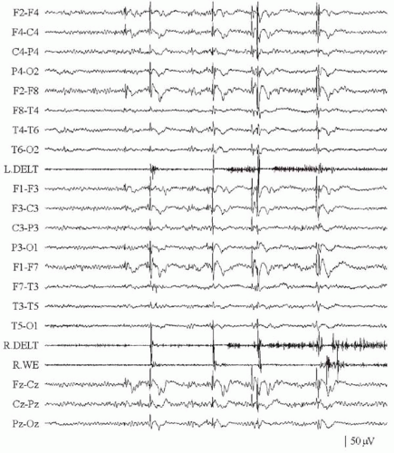
The electroencephalogram shows bursts of spike/poly-spike-and-wave complexes at 2 to 4 Hz. The muscle contraction responsible for the myoclonic jerk is usually followed by an EMG silence lasting up to 500 milliseconds (Fig. 26.2). The silent period is sometimes without a clear preceding jerk, although a mild contraction in nonsampled muscles is difficult to exclude. Bilateral synchronous EEG discharges and synchronous jerks in muscles from both sides of the body indicate a primary generalized myoclonus (50). Some patients can present with pure atonic seizures (46). Generalized tonic-clonic attacks and atypical absences usually occur as well.

Nonconvulsive status manifests as episodes of somnolence, stupor, apathy, or mild obtundation with drooling, associated with erratic muscle twitching and head nods. Beginning insidiously and progressing, such episodes may last from hours to days or even weeks, often with fluctuations, and have been considered indicative of a poor prognosis (43). In our experience, however, this is not necessarily the case. Nonconvulsive status is often accompanied by long runs of slow waves or spike-wave complexes so severely disorganized as to simulate a hypsarrhythmic pattern (35).

Tonic seizures, considered absent by some authors (6,51), have been found in up to 38% of cases in some series (43). Their presence does not necessarily predict an unfavorable outcome. “Vibratory” tonic seizures have also been mentioned (6,43).

The interictal electroencephalogram may be normal at onset (52). Bursts of 3-Hz spike waves may occur without apparent clinical manifestations and could be activated by sleep. The most suggestive findings are 4- to 7-Hz θ rhythms
with parietal accentuation and occipital 4-Hz rhythms, constantly blocked by eye opening (6). Variable lateralization of paroxysmal bursts is possible, although a consistently localized focus is unusual (50). A subset of patients are photosensitive.


Figure 26.2 Surface electroencephalographic (EEG)-electromyographic (EMG) polygraphic recording of a 13-year-old boy with myoclonicastatic epilepsy. Generalized myoclonic jerks are visible as high-amplitude myoclonic potentials on the EMG channels accompanied by a diffuse discharge of spike-wave complexes. A clear time-locked correlation is observed between the EEG spikes and the myoclonic EMG potentials. L.DELT = left deltoid muscle; R.DELT = right deltoid muscle; R.WE = right wrist extensor.


Course and Outcome

Despite the frequent seizure types, MAE is often self-limited and the seizures abate within 3 years in 50% to 89% of patients (6,43). Up to 58% of patients had normal intelligence quotients, while 20% had mild and 22% had severe mental retardation (6). Some children have mild behavioral problems, especially hyperactivity (43); others have intractable epilepsy. Pathophysiologic predictors of the outcome are poorly understood. The association between a poor mental outcome and frequent, especially prolonged, episodes of nonconvulsive status has been emphasized (12). Atypical absences, repeated generalized tonic-clonic seizures, and frequent falls have also been variably linked to a less favorable outcome. Nocturnal tonic seizures have been considered a bad prognostic indicator in some, but not all, reported series (43,52).


Differential Diagnosis

The progressive myoclonic epilepsies such as myoclonus epilepsy with ragged-red fibers (MERRF), Unverricht-Lundborg disease, and late-infantile neuronal ceroid lipofuscinoses (NCL) can be confused with MAE at onset, but later appearance of neurologic signs and continuous, multifocal myoclonus is usually sufficient for diagnosis. Late-onset cryptogenic epileptic spasms (53) can mimic myoclonic-astatic seizures, presenting as multiple daily episodes of violent falls; however, spasms tend to cluster in series with a typical periodicity, and the EEG recording shows different ictal, and usually interictal, features. Differentiation of MAE and Lennox-Gastaut syndrome is usually straightforward. The tonic and atonic attacks with slow spike-wave complexes is more consistent with the latter
but not incompatible with MAE. Other childhood epilepsies with predominant myoclonic attacks remain difficult to classify and in some cases may belong to the spectrum of MAE. It is difficult to subsume all myoclonic patients into single, well-defined syndromic entities (51). Of more use is to consider idiopathic epilepsies with prominent myoclonic seizures, such as BMEI, MAE, and other unclassifiable cases, as part of a continuum with different degrees of severity.


Treatment of Benign Myoclonic Epilepsy of Infancy and Myoclonic-Astatic Epilepsy

Treatment of myoclonic seizures is primarily with sodium valproate, ethosuximide, and the benzodiazepines. Lamotrigine does not seem to be effective but may be useful against generalized seizures, especially in MAE (54).

Patients with only myoclonic seizures often respond dramatically to regular doses of either valproate or ethosuximide. Ethosuximide produced a “good response rate” in 64% of patients (6); others may require higher doses of valproate (41). The combination is worth trying even when monotherapy with either drug has failed. Clonazepam may be effective but limited by behavioral side effects. Anecdotal reports (55,56) have championed topiramate, levetiracetam, acetazolamide, methsuximide, and sulthiame.

Resistant myoclonic-astatic seizures that cause disabling falls pose a therapeutic challenge. Uncontrolled trials of steroids or corticotropin have had limited success. Management should include practical measures, such as wearing a helmet, and considerable psychosocial support. The ketogenic diet may be effective but is difficult to maintain for long periods (6). Surgery, including callosotomy, is not generally indicated.


Severe Myoclonic Epilepsy of Infancy or Dravet Syndrome

Including SMEI with the myoclonic epilepsies is controversial, as myoclonus, although present in most children, can be transient and is not this syndrome’s main characteristic. Moreover, a subgroup of patients with SMEI do not present with myoclonic seizures at all (5,57). SMEI is characterized by multiple seizure types, including myoclonus, intractability despite treatment, and unfavorable evolution. It represents the prototype of an epileptic encephalopathy in which seizures are thought to be responsible for the deterioration of cerebral functions (58). Prolonged, often lateralized febrile seizures at onset are the most distinctive feature.


Etiology and Epidemiology

Although SMEI has a calculated incidence of 0.5 to 1 per 40,000 children (59,60), it represents 3% to 5% of all epilepsies starting in the first year of life (60) and 6.1% to 8.2% of those starting in the first 3 years of life (5,29). A 2:1 male preponderance is reported (60).

Genetic factors play a major pathogenetic role. A family history of febrile seizures or epilepsy is reported in 25% to 71% of patients (59,61), and affected monozygotic twins are on record (61,62). A link between SMEI and GEFS+ had been identified in several families (63). GEFS+ is associated with mutations of the SCN1A gene in approximately 10% of families (64); a study of seven children with SMEI found de novo truncating mutations of SCN1A in all of them (20). Similar results were subsequently confirmed (65). According to a genetic study of 93 patients with SMEI, SCN1A mutations are observed in approximately 35% of patients; in 10% of cases, mutations are inherited from asymptomatic or mildly affected parents (66). These data suggest a genetic heterogeneity with the possible involvement of a second gene, yet to be identified.


Clinical and Electroencephalographic Features

Frequent, usually (70%) febrile seizures, lasting several minutes to more than 1 hour, begin between age 2 months and the end of the first year of life in a normal child (5,67). Triggering factors are fever, infectious episodes without overt fever, and hot water immersion (5). Febrile seizures recur with a mean latency of 6 weeks between the first and second events, while afebrile seizures occur later (5-month mean latency from the first febrile seizure) (61). In the second and third years of life, myoclonic and atypical absences appear, but convulsive seizures remain a constant feature.

According to a detailed video-EEG description (5), generalized seizures are clinically and electrographically similar to those of idiopathic epilepsies, but the initial tonic phase can acquire a vibratory appearance, with high-frequency clonic activity. Unilateral clonic seizures, switching from side to side in different events or occasionally during the same seizure, are frequent at onset but rare after age 3 years. Postictal paresis is common, and EEG ictal activity is highly asymmetric. The so-called falsely generalized seizures are bilateral, tonic, and asymmetric followed by a clonic phase that is highly asymmetric in distribution of involved body segments and frequency of muscle activity. Ictally, electrodecremental activity is followed by slow spike-and-wave discharges, bilateral from onset, with variable asymmetry. In the classically similar “unstable” seizures, however, EEG ictal activity seems to migrate from one area of the brain to another during a single event.

Myoclonic seizures may appear between 1 and 5 years of age as massive, generalized myoclonic jerks, involving the axial muscles and causing falls to the ground. Highly variable in intensity, the jerks can present as nodding of the head, shrugging of shoulders, or abduction of the arms. Jerks can be isolated or grouped in brief clusters and can occur frequently during the entire day or concentrate on awakening, but tend to disappear during sleep (5). Polygraphic recordings show a generalized polyspike-and-wave discharge, time-locked to a brief EMG burst. These seizures appear to be generalized events; however, on EEG-EMG latency analysis, they are seen to spread contralaterally from a focal cortical activity in the
motor cortex of one hemisphere and either hemisphere can be activated in subsequent discharges (1). Multifocal erratic myoclonic jerks can involve the hands and face (5). Their origin remains speculative, as back-averaging studies did not produce any cortical transient time-locked to the jerks (1).

Atypical absences accompanied by head nodding or loss of postural control occur in 60% of patients (5) either between ages 1 and 3 years, concomitantly with myoclonic seizures, or between ages 5 and 12 years. EEG recordings show generalized, irregular 2- to 3.5-Hz spike-wave discharges lasting 3 to 10 seconds.

Nonconvulsive status epilepticus has been reported as fluctuating episodes of obtundation, with distal jerks, unsteadiness, and dribbling lasting from hours to several days (5).

Approximately 45% to 80% of patients experience focal seizures, either simple partial motor or complex partial, as early as 4 months or until 4 years of age (61). Prominent autonomic phenomena accompany complex partial seizures (5,67). Focal seizures may evolve to generalized or unilateral seizures.

Only gold members can continue reading. Log In or Register to continue

Stay updated, free articles. Join our Telegram channel

Oct 17, 2016 | Posted by in NEUROLOGY | Comments Off on The Myoclonic Epilepsies

Full access? Get Clinical Tree

Get Clinical Tree app for offline access