Treatment of Myoclonic Epilepsies of Childhood, Adolescence, and Adulthood


Treatment of Myoclonic Epilepsies of Childhood, Adolescence, and Adulthood


Marco T. Medina*

Iris E. Martínez-Juárez

Reyna M. Durón*

Pierre Genton

Renzo Guerrini§

Charlotte Dravet

Michelle Bureau

Katerina Tanya Perez-Gosiengfiao

Claudia Amador*

Julia N. Bailey

Franz Chaves-Sell||

Antonio V. Delgado-Escueta


*National Autonomous University of Honduras, Tegucigalpa, Honduras, California Comprehensive Epilepsy Program, David Geffen School of Medicine at UCLA, Los Angeles, California

§Epilepsy, Neurophysiology, and Neurogenetics Unit, Department of Child Neurology and Psychiatry, University of Pisa and Research Institute ‘Stella Maris’ Foundation, Pisa, Itlay

Centre Saint-Paul-Hôpital Henri Gastaut, Marseille, France

*National Autonomous University of Honduras, Tegucigalpa, Honduras

California Comprehensive Epilepsy Program, David Geffen School of Medicine at UCLA, Los Angeles, California

||Hospital Clinica Biblica, San José, Costa Rica


Introduction

Myoclonic epilepsies are still often misdiagnosed, treated with undue delay, or treated with inappropriate drugs. Difficulties in choosing the right treatment in these epilepsies have been discussed in recent reviews (1,2,3). New antiepileptic agents have opened new possibilities, but have also increased risks of paradoxical aggravation of myoclonic epilepsies. Specific treatment strategies of the different forms of idiopathic myoclonic epilepsies have been discussed in various chapters of this book and the reader can refer to them. However, a summary of this complex field would be useful for the clinician.

No Class I study, which implies a controlled and randomized study, has been performed regarding the myoclonic epilepsies. Most of the literature is based on uncontrolled comparisons, such as groups of patients or case studies. Some randomized trials have been conducted that include myoclonic epilepsies together with other primary generalized epilepsies.

In this chapter, we first describe the results of a long-term follow-up in 222 patients with juvenile myoclonic epilepsy (JME), subdivided into its subsyndromes. We analyze the responses to antiepileptic drug (AED) treatment based on the constellation of seizure types, common trigger factors, seizure types that persist after antiepileptic drug treatment, frequency of breakthroughs, and side effects of antiepileptic, during more than 10 years of follow-up. We next describe AEDs reported in literature used in the management of myoclonic absence epilepsy, eyelid myoclonic epilepsy, JME and familial adult myoclonic epilepsy (FAME). Finally, we provide an algorithm for treatment of JME based on our 222 patients and the Marseille/Nice cohort on JME. This algorithm also considers opinions of experts and available information from recent published studies.


Juvenile Myoclonic Epilepsy Consortium of GENESS: Treatment Results and Long-Term Outcome in 222 Patients


Classic Juvenile Myoclonic Epilepsy

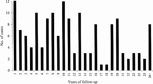
At the time we initially evaluated 161 classic JME patients (91F:60M), 60% (96/161) had myoclonic and tonic–clonic seizures, 34% (54/161) had myoclonic, tonic–clonic, and spanioleptic absence seizures, 6% (10/161) had myoclonic seizures only, and 1% (1/161) had both myoclonic and absence seizures. We were able to follow these 161 patients for a mean period of 11.6 years (range 1 to 41 years) (Fig. 25-1); 57% were female (91/161) and 43% were male (70/161).


FIG. 25-1. Years of follow-up in 161 patients with classic JME.

In evaluating the results of this long-term follow-up, we first asked how many patients were completely free of tonic–clonic grand-mal convulsions (Table 25-1). Eighty-five percent (137/161) were free from tonic–clonic seizures, most often because of AED treatment. We then asked how many patients were completely free of all type of seizures due to AED treatment. Of the 161 patients, 72 (54%) were seizure-free and did not suffer any breakthrough seizures during the follow-up period of 10.1 years (range 1 to 41 years).

We also asked how many patients had uncontrolled seizures in spite of treatment and the reason why. At the initial portion of the follow-up period, there were 89/161 patients who identified various trigger factors as responsible for breakthrough seizures (
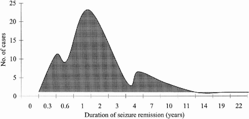
Table 25-2). In subsequent months and years of follow-up, some patients were able to correct trigger factors, such as sleep deprivation and noncompliance, thereby increasing the number of patients whose seizures completely ceased from 72 to 93. When analyzed separately, these 93 patients achieved complete freedom from seizures for a mean period of 34 months. Among them were 17 patients who were in remission from seizures for 4 to 11 years (Fig. 25-2).


TABLE 25-1. Long-Term Follow-Up and Outcome of Juvenile Myoclonic Epilepsy Subsyndromes


Classic JME


CAE evolving to JME


JME with adolescent pyknolepsy absence


JME with myoclonic astatic


Grand mal controlled


137/161 (85%)


26/35 (74%)


17/18 (94.4%)


6/8 (75%)


Persisting myoclonic seizures


31/161 (19%)


4/35 (11%)


2/18 (11%)


0


Persisting MS + ABS


5/161 (3%)


0


5/18 (28%)


0


Persisting grand mal


9/161 (5.5%)


6/35 (17%)


1/18 (6%)


1/8 (12.5%)


Persisting ABS only


0


16/35 (46%)


0


0


Persisting myoclonic astatic


0


0


0


1/8 (12.5%)


MS, myoclonic seizures; ABS, absence seizures.


TABLE 25-2. Trigger Factors for Seizures (Mainly Myoclonic Seizures) in 161 Patients with Classic JME


Trigger factors


No. of cases


%


Sleep deprivation


71


44


Awakening


61


38


Stress


32


20


Alcohol


26


16


Noncompliance


23


15


Menses


17


10


Fatigue


16


10


Light (TV, video games, strobe)


14


9


Physical activity


3


3


Cognitive tasks (e.g., reading)


3


3


Anger


1


1


Hunger (hypoglycemia)


1


1


FIG. 25-2. Remission of seizures (years) in 93 patients with classic JME.

Next, we turned our attention to the 68 patients who habitually suffered breakthrough seizures in spite of recognizing the responsible trigger factors (Table 25-2). Thirty-one of these patients only had myoclonic seizures more often on awakening. Five other patients had myoclonic and absence seizures, while 9 had breakthrough tonic–clonic grand-mal convulsions.

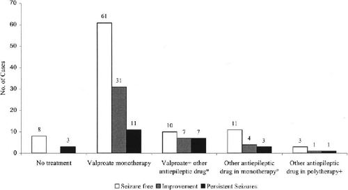
We next asked what AEDs patients were receiving, how long the patients remained seizure-free, and what were the adverse effects of AEDs. Of the 93 patients who were completely seizure-free, 85 were receiving AEDs. 60 patients (65%) were on valproate (VPA) monotherapy. They were seizure-free as long as 22 years, the mean interval of seizure-free period being 36 months.

Of seizure-free patients 11% (10/93) were receiving VPA plus one or more other AEDs. Some 15% (14/93) were taking AEDs other than VPA either as mono- or polytherapy.

Interestingly, there were eight patients (9%) who were seizure-free, but were not on any AEDs (Fig. 25-3). Among them was one patient who had been seizure-free for 11 years.


FIG. 25-3. Response to treatment according to AED regimen in 161 patients with classic JME. Seizure-free means no myoclonic, tonic–clonic, or absence seizures. Improvement means either 50% reduction in seizures or the persistence of breakthrough myoclonic or rare tonic–clonic seizures when trigger factors appear. Persistent seizures means no change in seizure pattern and frequencies. *, Other AEDs include: carbamazepine (CBZ), phenytoin (PHT), lamotrigine (LTG), clonazepam (CZP), ethosuximide (ESM), methsuximide (MSM), and lorazepam. One patient was seizure-free on valproate (VPA) + carbamazepine (CBZ), one patient on valproate (VPA) + phenytoin (PHT), five patients on valproate (VPA) + lamotrigine (LTG), one patient on valproate (VPA) + clonazepam (CZP), one patient on valproate (VPA) + topiramate (TPM), and one patient on valproate (VPA) + lorazepam. °, Other AEDs in monotherapy included lamotrigine (LTG), carbamazepine (CBZ), topiramate (TPM), phenobarbital (PB), primidone (PRM), and levetiracetam (LEV). Three patients were seizure-free on carbamazepine (CBZ), two patients on lamotrigine (LTG), two patients on phenobarbital (PB), two patients on levetiracetam (LEV), one patient on topiramate (TPM), and one patient on primidone (PRM). +, One patient was seizure-free on topiramate (TPM) + lamotrigine (LTG) and one patient on phenobarbital (PB) + clonazepam (CZ).

The most common side effects in 150 patients taking AEDs were weight gain (12 patients), tremors (6 patients), hair loss (5 patients), nausea and vomiting (3 patients), polycystic ovary (2 patients), gastritis (1 patient), diarrhea (1 patient), hepatotoxicity (1 patient) and memory problems (1 patient). All side effects were observed in the group of patients taking VPA either as mono- or polytherapy. Tremor was found in one patient taking topiramate (TPM) monotherapy. Weight gain was reported also in one patient taking primidone (PRM) monotherapy.

It seemed that the more combinations of seizure types, the harder to control seizures with AEDs. Among patients presenting with myoclonic seizures only, 80% (8/10) were seizure-free. Of patients presenting myoclonic and tonic–clonic seizures, 63% (60/96) were seizure-free. On the other hand, only 46% (25/54) of patients with myoclonic, tonic–clonic, and absence seizures were seizure-free. The sole patient who had myoclonic and absence seizures had not achieved seizure control.

Among 68 patients who had recurrent seizures, 3 patients chose not to take any medication. Two were planning to get pregnant; they suffered mainly from myoclonic seizures and rare tonic–clonic convulsions. Among these 68 patients, 25 reported dissatisfaction with AED treatment because seizure frequency did not change.

Episodes of convulsive tonic–clonic status epilepticus (two patients) and myoclonic and absence status (one patient) were uncommon and were reported in only three patients. We did not observe any significant difference regarding response to treatment among women and men. Of women, 57 were seizure-free (61%), compared to 36 men (52%) who were seizure-free.

We also reviewed what AEDs patients were receiving at the time they were initially referred to our service. Among the 161 classic JME patients, 104 (64%) received carbamazepine (CBZ) and/or phenytoin (PHT) prior to referral to our service. Only 14% reported some improvement of seizures. In contrast, 14 of 21 (66%) blamed CBZ monotherapy for increasing frequencies of tonic–clonic grand mal, absence, and myoclonic seizures. PHT in combination with VPA phenobarbital (PB) or CBZ increased seizure frequency even more [18/23 or 78% of patients receiving PHT]. PHT monotherapy also aggravated seizures of nine out of 21 patients (42%). PHT in combination with VPA, PB or PRM aggravated seizures in fewer patients (8/25 or 32%). Overall, CBZ and/or PHT increased seizure frequency in 50 of 104 patients (48%). In two patients, this drug combination could have triggered the first appearance of absence seizures. Genton et al. (1) described similar results. (Table 25-3)


TABLE 25-3. Response to Treatment with Traditional AEDs in Patients with Myoclonic Epilepsies of Adolescence


Patients from Marseille/Nice (n = 40)


Patients from GENESS consortium (n = 104)


Treatment


No.


Aggravated


No change


Improved


No.


Aggravated


No change


Improved


CBZ


Monotherapy


13


10


1


2


21


14


5


2


+ PHT


12


10


2


+ VPA


5


1


3


1


3


2


1


+ PB


6


5


1


+ PB + VPA


1


1


+ PHT + VPA


1


1


+ PHT + PB


1


1


8


6


2


+ PB + VGB + CLB


1


1


Without PHT, n (%)


26


17 (65)


5 (20)


4 (15)


24


16 (67)


6 (25)


2 (8)


All, including those treated in polytherapy with PHT n (%)


28


19 (68)


5 (18)


4 (14)


44


32 (73)


10 (22)


2 (5)


PHT


Monotherapy


4


1


2


1


21


9


8


4


+ VPA


1


1


1


6


2


3


1


+ PB


7


2


4


1


12


5


6


1


+ PRM


3


1


2


+ CLB


1


1


+ PB + VPA


1


1


+ PB + CLZ


1


1


+ CLZ


2


2


+ ZNS


1


1


Without CBZ, n (%)


14


4 (29)


8 (57)


2 (14)


46


17 (37)


22 (48)


7 (15)


All, including those treated in polytherapy with CBZ n (%)


16


6 (38)


8 (50)


2 (12)


66


33 (50)


26 (39)


7 (11)


PB or PRMa


Monotherapy


12


1


7


4


+ CLZ


2


2


+ CBZ


7


6


1


+ PHT


8


2


5


1


13


5


7


1


+ PHT + CBZ


1


1


8


6


2


All with PB/PRM alone or in polytherapy


16


9 (56)


6 (38)


1 (6)


35


12 (34)


16 (46)


7 (20)


Total patients, n (%)


40


23 (57)


11 (28)


6 (15)


104


50 (48)


39 (38)


15 (14)


Data taken from 40/172 patients from Marseille/Nice (23%) and 104/254 (41%) patients from the GENESS Consortium.


a Only 3 with PRM in the GENESS group.


Childhood Absence Epilepsy Evolving to Juvenile Myoclonic Epilepsy

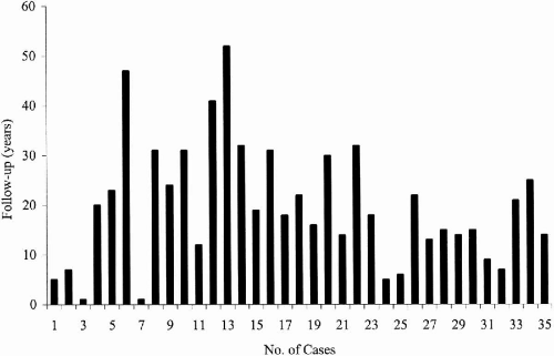
We followed 35 patients with childhood absence epilepsy (CAE) evolving to juvenile myoclonic epilepsy (JME) for a mean period of 19.8 years (range 1 to 47 years) (Fig. 25-4). When initially seen, 32 had pyknoleptic absences, tonic–clonic and myoclonic seizure, and only one patient had absence and myoclonic seizures only; 66% were female (23/35) and 44% were male (12/35).


FIG. 25-4. Long-term follow-up in a cohort of 35 patients with CAE evolving to JME.

During the long period of follow-up, 3 patients among the 35 original cohort achieved complete seizure control from 2 to 40 years (Table 25-1). Even though 32 patients had persisting seizures, VPA mono- (27 patients) or polytherapy [5 patients: combination with lamotrigine (LTG) or PB or zonisamide (ZNS) with levetiracetam (LEV)] completely suppressed tonic–clonic seizures in 23 patients (72%). Thus 26 patients out of a total number of 35, had satisfactory seizure control because grand-mal seizures had ceased. In this syndrome, absence seizures comprised the most common phenotype that persisted (16/35 patients). Rarely, breakthrough tonic–clonic (6/35 patients) and myoclonic seizures (4/35 patients) were observed.

Of three patients (3/35) who were completely free of seizures, one was taking PB and mephenytoin, another was on LEV and TPM, while one was on VPA monotherapy.


Juvenile Myoclonic Epilepsy with Adolescent-Onset Pyknoleptic Absence

We followed 18 patients with adolescent-onset pyknoleptic absence mixed with JME for a mean period of 13.4 years (range 5 to 26 years). A majority of patients were female (13/18 or 72%). Myoclonic, tonic–clonic, and absence seizures were present in 15/18, while 3/18 had myoclonic and absence seizures only.

Ten patients (56%) were seizure-free, while eight (44%) still had seizures. However, 7 of these 8 patients had no persisting tonic–clonic seizures, bringing a total of 17 out of 18 patients who were satisfied with seizure control because grand-mal convulsions stopped. The mean time that patients were seizure-free was 33 months. Characteristically, absences with or without myoclonic seizures persisted in 5/8. Myoclonic seizures persisted in 2/8 and tonic–clonic in one.

Seventy percent (7/10) were seizure-free on VPA monotherapy while 30% (3/10) were seizure-free on VPA associated with LTG, TPM, or LEV. Seven patients decreased seizure frequency, but in one, seizures persisted.

Side effects associated with AEDs were weight gain (three patients), depression (one patient), hair loss (one patient), hirsutism (one patient), and tremor (one patient).

PHT, CZP, PB, PRM, and ZNS had been previously used in 11/18 (61%) of the probands, but were changed due to increase in myoclonic, tonic–clonic, and absence seizures. In one patient, absence seizures appeared at 15 years of age while taking PHT and CBZ.


Juvenile Myoclonic Epilepsy with Astatic Seizures during Adolescence

Eight patients who had astatic seizures during adolescence were followed up for a mean period of 11.1 years (range 3–18 years). Four were female and four men. Myoclonias with or without tonic–clonic and astatic seizures were present in all patients; only one reported spanioleptic absences.

Six patients were seizure-free (6/8). One had persistent astatic seizures, while another one had breakthrough tonic–clonic seizures when carbamazepine (CBZ) levels decreased. Mean time without seizures was 20 months.

Trigger factors for seizures were awakening (four patients), noncompliance (three patients), fatigue (two patients), alcohol (one patient), light (one patient), menses (one patient), sleep deprivation (one patient), and monetary problems leading to noncompliance (one patient).

Four out of the seven patients that were seizure-free were on VPA monotherapy; of the other two, one was taking VPA plus CBZ the other was on VPA plus LTG. Reported side effects were weight gain (two patients), hair loss (one patient), and tremor (one patient).

In one patient, the previous use of CBZ and PHT was related to the appearance of the myoclonic-astatic seizures. One patient previously treated with PHT had daily astatic seizures; this patient remained seizure-free after VPA monotherapy was started. Finally, one patient with PHT and PRM had persistent tonic–clonic seizures that stopped with VPA monotherapy.


Treatment of Specific Myoclonic Epilepsies According to Literature (Tables 25-4, 25-5 and 25-6)


TABLE 25-4. Commonly used antiepileptic drugs in myoclonic epilepsies


Antiepileptic drugs (AEDs)


Usual adult daily dosesa


Usual child daily dosesa


Time required to reach steady state (days)


Blood levelb


Elimination half-life (h)c


Protein binding (%)


Side effects


First- and second-generation AEDs


Clobazam (CLB)d


10–30 mg (divided)


2.5 mg/kg/day


Not established


10–30


85


Sedation, ataxia, behavioral changes, increase salivation


Clonazepam (CZP)


1–10 mg (divided)


0.1–0.2 mg/kg/day


10–70 ng/ml


20–40


45


Sedation, ataxia, behavioral changes, increase salivation


Ethosuximide (ESM)


1000 mg (divided)


20–30 mg/kg/day


7–10


50–100 µg/ml


20–60


None


Nausea, fatigue, gastrointestinal irritation, behavioral changes


Methsuximide (MSM)


300–1200 mg (divided)


20 mg/kg/day


8–16


10–40 mg/l


30–80


Low


Somnolence, ataxia, headache, behavioral changes, nausea, vomiting


Valproate (VPA)


1000–3000 mg (divided)


15–60 mg/kg/day


1–2


40–150 µg/ml


6–15


80–95


Nausea, vomiting, somnolence, weight gain, gastrointestinal irritation, tremor, transient alopecia, hepatotoxicity, thrombocytopenia, hyperammonemia


Third generation AEDs


Lamotrigine (LTG)


100–500 mg (divided)


2–8 mg/kg/day (monotherapy) 1–5 mg/kg/day (polytherapy)


7–10


1–10 µg/ml


29(14–60)


55


Headache, nausea, dizziness, diplopia, rash, ataxia, Stevens-Johnson syndrome, toxic epidermal necrolysis


Levetiracetam (LEV)


1000–3000 mg (divided)


40 mg/kg/day


2


5–40 mg/l


6–8


None


Behavioral changes


Topiramate (TPM)


100–400 mg (divided)


6–15 mg/kg/day


5


4–10 mg/l


19–23


9–17


Lethargy, paresthesias, ataxia, poor concentration, renal lithiasis, acuteangle closure glaucoma, weight loss


Zonisamide (ZNS)


200–400 mg (divided)


4–20 mg/kg/day


14


15–40 mg/l


50–70


40–60


Fatigue, somnolence, behavioral changes, anorexia, renal lithiasis


a This is a maintenance dose; a lower dose is necessary when initiating therapy.

b Therapeutic levels may change with multiple drugs.

c Half-life may also change with multiple drugs. Half-life is given for adult dosages. Steady state is reached in four half-lives.

d Not FDA approved.

Only gold members can continue reading. Log In or Register to continue

Stay updated, free articles. Join our Telegram channel

Jul 27, 2016 | Posted by in NEUROLOGY | Comments Off on Treatment of Myoclonic Epilepsies of Childhood, Adolescence, and Adulthood

Full access? Get Clinical Tree

Get Clinical Tree app for offline access