49 Viral Diseases
Herpes Simplex Encephalitis
Clinical Vignette
An independent 74-year-old man left a family wedding reception early because he did not feel well; he complained of mild nausea and general malaise. His daughter called the next day and when he did not answer the phone she went to his home to check on him, discovering him wandering in his backyard acutely confused. She convinced him to go to the emergency department, where he was found to be febrile with a temperature of 38.5° C (101.3° F). He soon became unresponsive to verbal stimuli, had conjugate right eye deviation, neck stiffness, and bilateral palmar grasps. He withdrew to noxious stimuli; plantar responses were flexor.
A noncontrast head computed tomography (CT) was unremarkable. Cerebrospinal fluid (CSF) examination demonstrated a WBC count of 45/mm3, predominantly lymphocytes, protein of 110 mg/dL, and a normal glucose level. Intravenous acyclovir 10 mg/kg every 8 hours was begun. A magnetic resonance image (MRI) of the brain demonstrated T2-weighted hyperintensity with edematous changes in the left insular cortex region of the inferior temporal lobe, the parahippocampal gyrus, and the hippocampus, extending into the subthalamic nucleus suggestive of herpes simplex virus (HSV) encephalitis. Electroencephalography (EEG) demonstrated periodic lateralized epileptiform discharges (PLEDs). This diagnosis was confirmed by HSV polymerase chain reaction (PCR) 4 days after symptom onset. The patient gradually improved and was treated with a 21-day course of acyclovir. After a short stay in a rehabilitation facility, he was discharged home.
Comment: This is a fine example of the rapidity with which herpes simplex encephalitis (HSE) will declare itself and the urgent need to consider the diagnosis in any acutely confused patient, initiating treatment based on clinical judgment alone without waiting for definitive diagnostic proof to become available. Unless this type of decision making takes place, the HSE will have caused irreversible cerebral damage, particularly involving the temporal lobes with their memory and language function modalities.
Etiology
A wide spectrum of viral agents may cause an infectious encephalitis. Diagnosis and management are dependent on identifying the precise causative agent. Only a few viruses are amenable to specific antiviral therapy. Therefore, prevention strategies are particularly important, especially for arthropod-borne viruses such as West Nile virus (WNV) and eastern equine encephalitis (EEE).
HSE is the most common acute encephalitis in the United States, with an annual incidence of 1 in 250,000 to 1 in 500,000. It affects all ages and both sexes equally, without significant seasonal variation. Early antiviral treatment significantly reduces mortality, but morbidity remains unacceptably high. Most cases of HSE are caused by oral herpes (herpes simplex virus [HSV] type 1); however, genital herpes (HSV-2) is more common among neonates with disseminated disease. HSE commonly develops as a recurrent infection but on occasion may occur during primary infection. Animal data verify the presence of retrograde virus transport into the brain via olfactory or trigeminal nerves. However, the human disease pathogenic pathways are not fully clarified. There is a predilection for HSE, once established, to lead to a hemorrhagic necrosis with inflammatory infiltrates and cells containing intranuclear inclusions.
Clinical Presentation
The symptoms and signs of patients with subacute or acute focal encephalitis are generally rather nonspecific, with fever, headache, and altered consciousness being the most common. Focal manifestations often include seizures, typically complex partial in character because of the temporal lobe’s predisposition to develop a herpes infection. It is common for these patients to also develop language difficulties, personality changes, hemiparesis, ataxia, cranial nerve defects, and papilledema (Fig. 49-1). The differential diagnosis includes stroke, brain tumors, other viral encephalitides, bacterial abscesses, tuberculosis, cryptococcal infections, and toxoplasmosis.
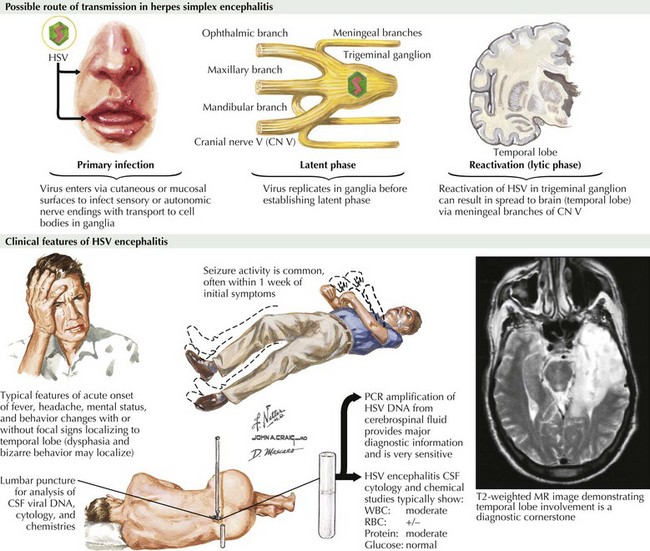
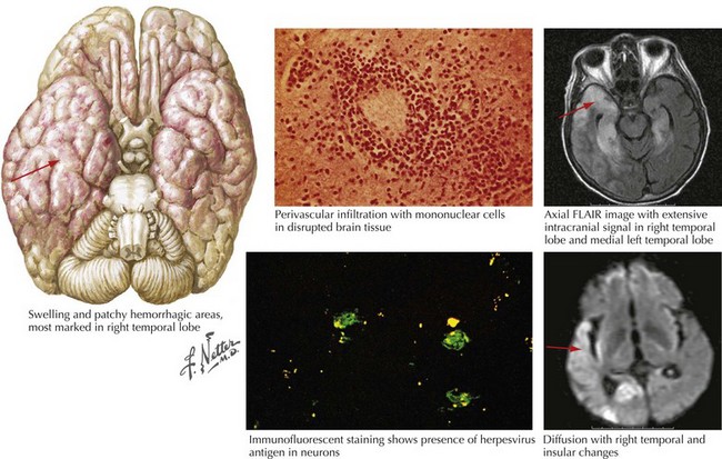
Diagnosis
One of the major issues in diagnosis is for the examining clinician to put HSE into his or her diagnostic spectrum very early on in the temporal profile of the patient’s illness. If this is not applied, a major therapeutic window allowing for successful treatment is sometimes lost. One of the saddest neurologic clinical scenarios is to evaluate a patient for confusion that has been present for the past 3–5 days and wrongly attributed to medication, or minor infection such as one involving the urinary or respiratory tracts. This is particularly liable to occur in a previously mentally vital senior citizen who develops a febrile illness with acute confusion and the change in mental status is presumed to be secondary to the fever per se, secondary nonspecific metabolic or toxic effects of empirical antibiotics, a stroke, or even “sundowning.”
For patients with suspected encephalitis, the initial diagnostic studies must include a CT scan (to rule out a mass effect) and then immediate CSF examination. CT scan results are abnormal in 50% of cases early on and usually demonstrate localized edema, low-density lesions, mass effects, contrast enhancements, or hemorrhage. MRI and EEG may be subsequently obtained for further confirmation. These usually demonstrate major temporal lobe damage (Fig. 49-2); however, a normal study does not exclude an HSE diagnosis. If such does occur, it is often wise to repeat the study within a few days, particularly if the patient continues to be confused.
CSF findings are nonspecific, often including a lymphocytic pleocytosis with a slight protein increase. Abnormal CSF findings are found in 96% of biopsy-proven HSE cases. EEG may show repetitive spiked, sharp wave discharges and slow waves localized to the involved area often as PLEDs.
The accuracy of PCR testing for HSV-DNA to detect HSV-1 and -2 in CSF compares favorably with the previous use of brain biopsy. This methodology provides excellent sensitivity and specificity (90–98%). The viral sequence for HSV may be detected months after the acute episode and may be negative in early disease phases. PCR should not be used to monitor therapy success. No standardized commercial assay is available. Brain biopsy was previously the gold standard for specificity, but it is rarely indicated now with the widespread availability of HSV PCR testing. If used, biopsy specimens are examined for both histopathologic changes and HSV antigens by immunofluorescence testing and appropriate culture techniques.
Therapy
Immediate initiation of acyclovir is indicated the moment HSE is clinically suspected. This relatively benign medication has the greatest chance of efficacy if it can be initiated very early on in the patient’s clinical course. The excellent outcome of the patient in the initial vignette in this chapter emphasizes the absolute importance for primary care and emergency medicine physicians to immediately consider HSE in individuals of any age who experience relatively acute changes in mental status. Unfortunately, if immediate therapy is not commenced at presentation, the outcome is usually poor, with minimal chance of return to independent living.
Prognosis
Before the availability of intravenous (IV) acyclovir, mortality from HSE was approximately 70%. If one is able to initiate antiviral therapy within the first 24 hours of symptom onset, the prognosis is much better for long-term outlook if the patient is fully treated for 21 days. This approach has reduced both mortality and morbidity substantially. Overall, although morbidity remains high, with 60–70% of patients having significant neurologic deficits, the mortality is now 10–20%.
On rare occasions, a patient seemingly doing well with initial therapy has a relapse. Inadequate early dosing of the acyclovir is the usual cause. Thus, initial treatment regimens must include daily doses of 30 mg/kg intravenously usually in three separate aliquots of 10 mg/kg.
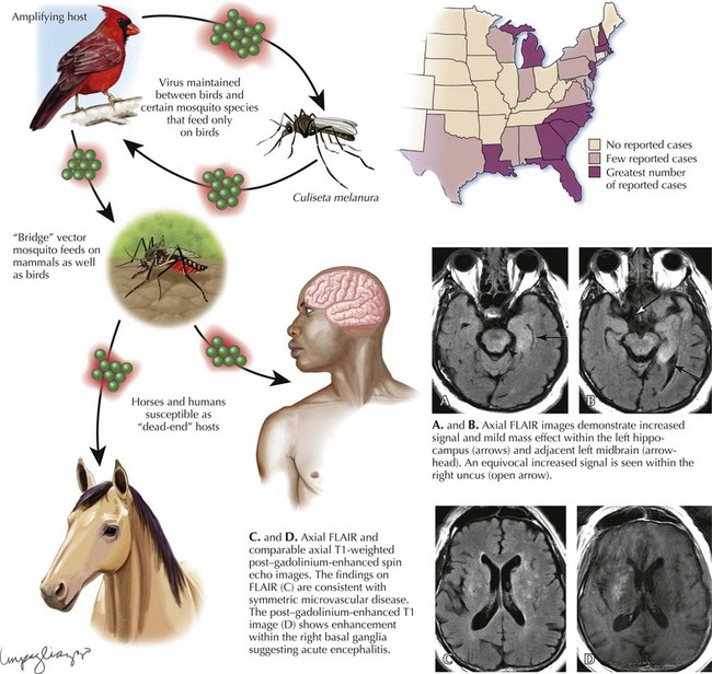
Eastern Equine Encephalitis
Clinical Vignette
A 60-year-old New Hampshire man presented in mid-August with 2 weeks of headache followed by dizziness and unsteady gait, then nausea and vomiting. He walked his black Labrador retriever past a local pond in the woods every day, sustaining multiple mosquito bites. He had type II diabetes mellitus and history of a prior nephrectomy for renal cell carcinoma. At clinical presentation he was febrile, 38.3° C (101° F), somnolent but arousable, was vague answering questions, there were fine tremors in his hands, and muscle stretch reflexes were globally depressed.
Brain MRI revealed increased signal with mild mass effect in the left hippocampus. Spinal fluid examination demonstrated a WBC of 1,860/mm3 (81% polymorphonuclear neutrophils (PMNs), 11% lymphocytes, 8% monocytes), 26 red blood cells/mm3, protein 106 mg/dL, glucose 98 mg/dL (serum 209), and Gram stain negative. He had continued fevers with progressive gait ataxia, upper extremity weakness, and memory loss. Bacterial cultures and studies for Borrelia burgdorferi, Treponema pallidum, herpes simplex virus, and West Nile virus were negative. CSF IgM and plaque assay were positive for eastern equine encephalitis. He gradually recovered motor function over the next few months with supportive care and 1 year later he had minimal residual cognitive deficits.
Epidemiology
Eastern equine encephalitis virus is a member of the family Togaviridae, genus alphavirus, that is found in the eastern half of the United States. Eastern equine encephalitis (EEE) is a mosquito-borne viral disease. Here it causes disease in humans, horses, and some bird species. It generally takes from 3 to 10 days to develop symptoms of EEE after being bitten by an infected mosquito. An average of 5 human cases occur per year (approximately 220 confirmed cases in the United States between 1964 and 2004, most frequently in Florida, Georgia, Massachusetts, and New Jersey). EEE virus transmission is most common in and around freshwater hardwood swamps in the Atlantic and Gulf Coast states and the Great Lakes region. The main EEE virus transmission cycle is between birds and mosquitoes.
Clinical Presentation and Treatment
Most persons infected with EEE virus do not demonstrate a clearly discernible illness. In those individuals who do develop clinical illness, symptoms range from mild flu-like illness to a fulminating encephalitis eventually leading to coma and death. The mortality rate from EEE is approximately 33%, making it one of the most deadly mosquito-borne diseases in the United States.
Diagnosis
Laboratory diagnosis of EEE virus infection is based on serology, especially IgM testing of serum and CSF, and neutralizing antibody testing of acute- and convalescent-phase serum. MRI is the most sensitive imaging modality for diagnosis of EEE (Fig. 49-3). The most commonly affected areas of the central nervous system (CNS) include the basal ganglia (unilateral or asymmetric, with occasional internal capsule involvement) and thalamic nuclei. Other areas include the brain stem (often the midbrain), periventricular white matter, and cortex (most often temporally). Affected areas appear as increased signal intensity on T2-weighted images.
Therapy/Prognosis
There is no specific treatment for EEE; optimal medical care includes intensive hospitalization and supportive care.
Approximately half of those persons who survive EEE will have mild to severe permanent neurologic damage, particularly involving cognitive impairment. Those older than age 50 and younger than age 15 years appear to be at greatest risk for developing severe EEE.
West Nile Virus
Etiology/Epidemiology
West Nile virus is a flavivirus usually found in Africa, West Asia, and the Middle East. The Middle Eastern strains are most closely related genetically to the St. Louis encephalitis virus found in the United States. There are many potential animal, ornithologic, and insect reservoirs including humans, horses, some other mammals, birds, and mosquitoes. The West Nile virus was not documented in the Western Hemisphere until 1999. In the temperate zones of the world, West Nile encephalitis cases occur primarily in the late summer or early autumn. In southern climates, where temperatures are milder, WNV can be transmitted year-round.
Clinical Presentation
West Nile fever is typically a mild disease characterized by flu-like symptoms that develop 3–15 days after the bite of an infected mosquito. West Nile fever usually lasts only a few days and does not seem to cause any long-term health effects. Mild fever, headache, body aches, occasional skin rash, and swollen glands are the most common symptoms.
However, there is a more severe disease spectrum that can manifest as encephalitis, meningitis, or meningoencephalitis. A poliomyelitis-like illness is also described, with an acute proximal and asymmetric flaccid paralysis, very occasionally occurring during recent outbreaks in the United States. Neurophysiologic, radiologic, and pathologic studies suggest that WNV has a proclivity to damage anterior horn cells within the spinal cord.
Human Immunodeficiency Virus (HIV)
Clinical Vignette
A 31-year-old mother of a 13-month-old child presented to the emergency department with headache, vertigo, diplopia, and an unsteady gait. She had been treated with antibiotics for acute sinusitis during the preceding 8 days. Her neurologic examination demonstrated a lethargic, restless, febrile woman with meningismus, photophobia, horizontal nystagmus, and slight appendicular ataxia of her right arm and leg.
Brain CT demonstrated diminished absorption bilaterally in both thalami and to a lesser degree her internal capsules, midbrain, pons, and right posterior temporal lobe. There were signs of a primary maxillary and sphenoid sinusitis. Spinal tap demonstrated a CSF with moderate increased pressure of 275 mm/CSF, a cell count of 485 white blood cells (84% neutrophils), a protein concentration of 106 mg/mL, and glucose of 66 mg/dL. Intravenous antibiotics as well as acyclovir were begun. Gram stain and culture were negative initially and on a repeat study within less than 1 day.
During the first day of admission, she developed increased obtundation and intermittently varied automatisms. Bilateral flexor posturing and intermittent left-sided extensor posturing developed during her second day of hospitalization. Dexamethasone was administered every 6 hours. EEG demonstrated bilateral 3–5-Hz activity. Repeat imaging demonstrated extension of the low-density lesions into the basal ganglia and frontal lobe operculum. Temporal lobe biopsy was negative for HSV virus. Three days after admission, all spontaneous movements ceased; her pupils became dilated, fixed, and nonreactive; she was now areflexic and did not respond to any form of sensory stimulation. EEGs were electrically silent on two occasions over the next 24 hours. She died on the fifth hospital day.
A history of marked sexual promiscuity became available during her hospitalization. CSF culture was eventually positive for HIV although no serum or CSF antibodies to HIV were defined; all other cultures for various microbes were negative. Pathologically, there was an encephalopathic demyelinating process affecting cerebral white matter, the thalamus, and brain stem with acute neuronal damage. There was no associated vasculitis.
Comment: This case, seen at Lahey in the mid-1980s, added further support to the proposal that HIV is a primary neurotropic virus. Our experience emphasized the importance of considering HIV in the differential diagnosis of any acute encephalitis even if the patient is HIV antibody negative. Here the initial antibody negativity supported the concept that this patient’s encephalitis represented the primary phase of her HIV. Today when one wishes to consider an acute HIV infection in the setting of a negative HIV antibody one now has available an HIV viral load study. This will be positive, despite a negative HIV antibody study, if the patient has an active HIV infection. Thus, one will not need to depend on a viral culture to make the diagnosis as occurred with this patient. Such was not available at the time we evaluated this person.

Full access? Get Clinical Tree

