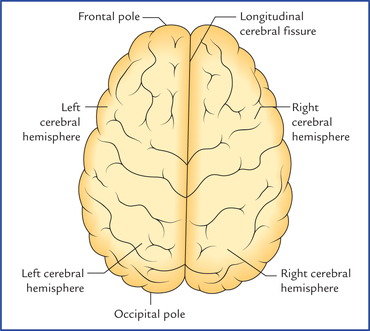
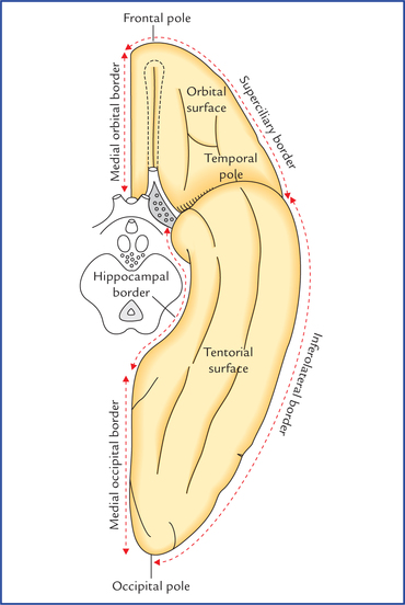
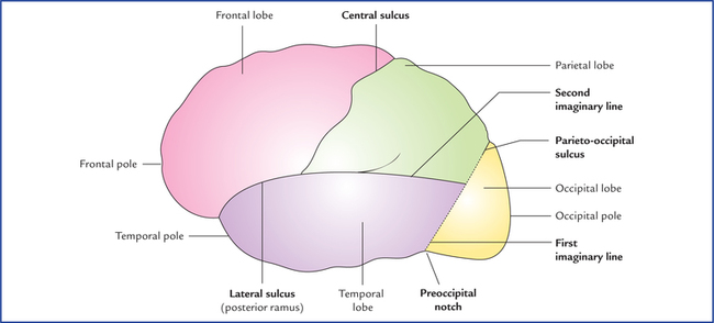
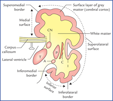
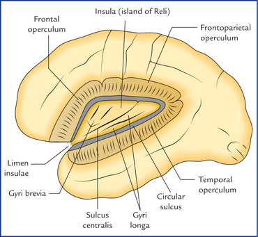
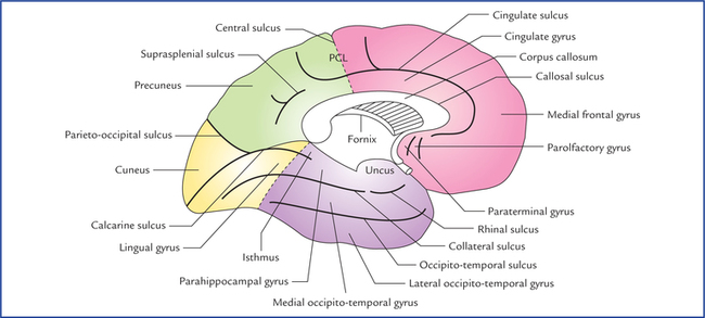
12 The cerebrum is the largest part of the human brain that fills most of the cranial cavity. Its large size is the result of a progressive (telencephalization) centralization of the various higher sensory and motor centres of the brain during evolution. The cerebrum is a heavily, convoluted bilobed structure (Fig. 12.1). The two lateral halves are called cerebral hemispheres. When the two cerebral, hemispheres are viewed together from above, they assume the shape of an ovoid mass, which is broader behind than in front. The widest transverse diameter corresponds with a line connecting the two parietal tuberosities. The longitudinal cerebral fissure is occupied by the following structures: 1. Falx cerebri (a sickle-shaped fold of dura mater). 2. Fold of arachnoid that follows the surfaces of the falx cerebri. 3. Pia mater covering the medial surface of the falx cerebri. 4. Anterior cerebral arteries and veins (which lie in the subarachnoid space between the arachnoid and the pia). Each cerebral hemisphere consists of: (a) an outer layer of grey matter called cerebral cortex, (b) an inner mass of white matter, (c) large masses of grey matter embedded in the basal part of the white matter called basal ganglia/basal nuclei, and (d) a cavity within it called lateral ventricle (Fig. 12.2). Fig. 12.2 Coronal section of the cerebral hemisphere showing its structure. Also note the borders and surfaces of the cerebral hemisphere. (CN = caudate nucleus, T = thalamus, P = putamen, C = claustrum, A = amygdaloid body.) The external features of the cerebral hemisphere include poles, surfaces, borders, sulci, and gyri. • The frontal pole at the anterior end of the hemisphere is more rounded than the occipital pole. It lies opposite the medial part of the superciliary arch. • The occipital pole at the posterior end of the hemisphere is more pointed than the frontal pole. It lies at a short distance superolateral to the external occipital protuberance. • The temporal pole between frontal and temporal poles points forwards. It fits into the anterior part of the middle cranial fossa and is overhung by the lesser wing of the sphenoid. Each cerebral hemisphere has three surfaces – superolat-eral, medial, and inferior (Fig. 12.2). 1. The superolateral surface is most convex and most extensive. It faces upwards and laterally and conforms to the corresponding half of the cranial vault. 2. The medial surface is flat and vertical. It presents a thick C-shaped cut surface of the corpus callosum. 3. The inferior surface is irregular to adopt the floors of anterior and middle cranial fossae. It is divided into two parts by a deep horizontal groove or sulcus, the stem of lateral sulcus, viz. (a) a small anterior part, the orbital surface, and (b) a large posterior part, the tento-rial surface. Each cerebral hemisphere presents six borders (Figs 12.2 and 12.3), viz. superomedial, superciliary, inferolateral, medial orbital, medial occipital and inferomedial. 1. The superomedial border separates the superolateral surface from the medial surface. 2. The superciliary border is at the junction of superolateral and orbital surfaces. It lies just behind the superciliary arch hence its name strictly speaking, it is the orbital part of the inferolateral border. 3. The inferolateral border separates the superolateral surface from the tentorial surface. Posteriorly this border exhibits a notch, the preoccipital notch about 3 cm in front of the occipital pole. This notch is used as a useful surface landmark. 4. The medial orbital border (Fig. 12.3) separates the medial surface from the orbital surface. 5. The inferomedial/hippocampal border (Fig. 12.3) surrounds the cerebral peduncle. It is formed by the medial aspect of the uncus and parahippocampal gyrus. 6. The medial occipital border (Fig. 12.3) separates the medial surface from the tentorial surface. To discuss further about sulci and gyri and other aspects of the cerebral hemisphere, the superolateral surface of the hemisphere is arbitrarily divided into four lobes – frontal, parietal, temporal and occipital with the help of: (a) three main sulci, central, lateral and parieto-occipital, and (b) two imaginary lines. The first imaginary line is a vertical line joining the parieto-occipital sulcus to the preoccipital notch, and the second line is a backward continuation of the horizontal part of the posterior ramus of the lateral sulcus till it joins the first line (Fig. 12.4). The parietal lobe lies behind the central sulcus and in front of the upper part of the first imaginary line. Below it is bounded by the posterior ramus of lateral sulcus and the second imaginary line. The insula is the submerged (hidden) portion of the cerebral cortex in the floor of the lateral sulcus (Fig. 12.5). It has been submerged from the surface during development of brain due to the overgrowth of the surrounding cortical areas and can be seen only when the lips of the lateral sulcus are widely pulled apart. It is triangular in shape and surrounded all around by a sulcus, the circular sulcus except anteroinferiorly at its apex called limen insulae which is continuous with the anterior perforated substance. Fig. 12.5 The insula (island of Reil) exposed by removing the opercula. Note: Insula is also called ‘central lobe.’ Fig. 12.6 Superolateral surface of the left cerebral hemisphere showing lobes, sulci and gyri. (SFG = superior frontal gyrus, MFG = middle frontal gyrus, IFG = inferior frontal gyrus (a. pars orbitalis, b. pars triangularis, c. pars opercularis), STG = superior temporal gyrus, MTG = middle temporal gyrus, ITG = inferior temporal gyrus, IPL = inferior parietal lobule.) The middle cerebral artery and deep middle cerebral vein lie on the surface of the insula. • The prefrontal sulcus often broken into two or three parts, runs downwards and forwards parallel and little anterior to the central sulcus. The area between the central and precentral sulci is called precentral gyrus. • Anterior to the precentral sulcus there are two sulci called superior and inferior frontal sulci which run horizontally. These sulci divide the region of frontal lobe in front of precentral sulcus into superior, middle, and inferior frontal gyri. • The anterior and ascending rami of lateral sulcus divide the inferior frontal gyrus into three parts. The part below the anterior ramus is called pars orbitalis, the part between the anterior and ascending rami the pars trian-gularis and the part posterior to the ascending ramus, the pars opercularis. • The postcentral sulcus runs downwards and forwards, a little behind and parallel to the central sulcus. The area between these two sulci is called the postcentral gyrus. • The rest of the parietal lobe is divided into a superior and inferior parietal lobules by an intraparietal sulcus which runs horizontally backwards from the postcentral sulcus. • The upturned posterior end of the posterior ramus of lateral sulcus, and the posterior ends of superior and inferior temporal sulci extends into the inferior parietal lobule to divide it into three parts: (a) the part that surrounds the posterior ramus of lateral sulcus is called supra marginal gyrus, (b) the part surrounding the superior temporal sulcus, the angular gyrus, and (c) the part surrounding the inferior temporal sulcus, the arcus temporo-occipitalis. 1. Lateral occipital sulcus runs horizontally and divides this lobe into superior and inferior occipital gyri. 2. Lunate sulcus is C-shaped sulcus with forward convexity just in front of the occipital pole. 3. Transverse occipital sulcus runs downwards into the uppermost part of the occipital lobe from the supero-medial border of hemisphere, a little behind the parieto-occipital sulcus. The presence of corpus callosum is the most conspicuous feature seen on the medial surface of the cerebral hemisphere. It is C-shaped thick bundle of commissural fibres. It consists of a central part, the trunk, a thick posterior end, the splenium and curved anterior end, the genu. Fig. 12.7 Medial surface of the left cerebral hemisphere showing lobes, sulci and gyri. (Note: In this figure tentorial surface of hemisphere is also seen.) The interrupted lines indicate the approximate boundaries of the lobes. (PCL = paracentral lobule.) The sulci and gyri on the medial surface are located above, in front and behind the corpus callosum. • Cingulate sulcus: It is the most prominent sulcus which follows a curved course about 1 cm above and parallel to the upper convex margin of the corpus callosum. Anteriorly it ends below the genu of corpus callosum, posteriorly it turns upwards to reach the superomedial border of the hemisphere a little behind the upper end of the central sulcus. The area between the cingulate sulcus and the corpus callosum is termed cingulate gyrus. • Just in front of lamina terminalis, there are paraterminal and parolfactory gyri and anterior and posterior parol-factory sulci. • Callosal sulcus. It separates the cingulate gyrus from the corpus callosum. • The part of medial surface between the cingulate sulcus and the superomedial border of hemisphere is divided by a short offshoot sulcus ascending from the cingulate sulcus above the middle of the trunk of corpus callosum into two parts: • The posterior part of medial surface behind the para-central lobule has two main sulci: the calcarine sulcus, and the parieto-occipital sulcus. (a) Calcarine sulcus. It is already described on page 142. A small region between the splenium and calcarine sulcus is termed isthmus. (b) Parieto-occipital sulcus. It is already described on page 142.
Cerebrum
External Features of the Cerebral Hemisphere
Poles (Figs 12.2 and 12.3)
Surfaces
Borders
Sulci and Gyri
Lobes of Cerebral Hemisphere (Fig. 12.4)
Insula/island of Reil (also called central lobe)
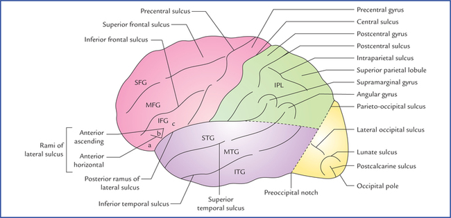
Sulci and Gyri on the Superolateral Surface of the Cerebral Hemisphere (Fig. 12.6)
In the frontal lobe
In the parietal lobe
In the occipital lobe
Sulci and Gyri on the Medial Surface of the Cerebral Hemisphere (Fig. 12.7)
Related posts:
![]()
Stay updated, free articles. Join our Telegram channel

Full access? Get Clinical Tree


Cerebrum