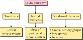
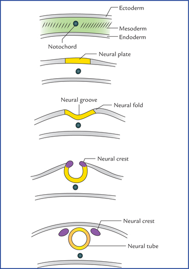
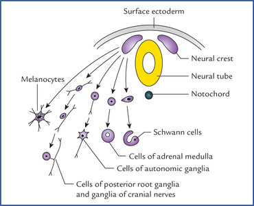
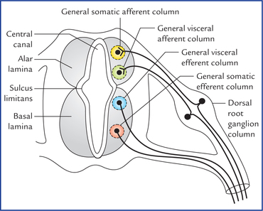
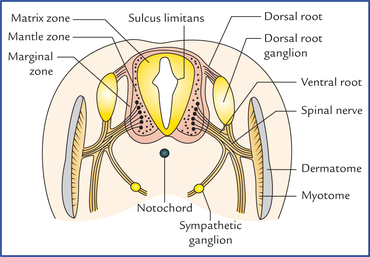
Development of the nervous system

1


Development of the Nervous System


A study of development of the nervous system helps to understand its complex organization and the occurrence of various congenital anomalies.


The whole of the nervous system is derived from ectoderm except its blood vessels and some neuroglial elements.


The specific cell population of the early ectoderm, which gives rise to entire nervous system and special sense organs is termed neural ectoderm. The neural ectoderm later differentiates into three structures: neural tube, neural crest cells, and ectodermal placodes. The neural tube gives rise to the central nervous system (CNS), the neural crest cells form nearly all the peripheral nervous system and ectoder-mal placodes contribute to the cranial sensory ganglia, hypophysis and inner ear (Flowchart 1.1).


Formation of Neural Tube (FIG. 1.1)


In the early embryonic disc, at about 16th day of embryonic life, the ectoderm overlying the newly formed notochord thickens in the midline forming the neural plate. As somatic mesoderm develops on either side of notochord, the margins of neural plate are elevated as neural folds, as a result the centre of the plate sinks, creating the neural groove. The neural folds gradually move together towards the mid-line and finally fuse to form a cylindrical neural tube that loses its connection with the surface ectoderm. The process of neural tube formation is termed neurulation.


The fusion of neural folds begins in the middle (region of fourth somite on 20th day of embryonic development) and it simultaneously proceeds in the cephalic and caudal directions. The fusion at the cranial and caudal ends of neural tube are somewhat delayed, forming small openings called anterior and posterior neuropores. The neural tube and surrounding amniotic cavity, therefore, remain temporarily in open communication with each other through these pores. The anterior neuropore closes in the middle of the 4th week at 18–20 somite stage (i.e. on 25th day) and posterior neuropore closes at the end of 4th week at about 25 somite stage. By the time the neural tube is completely closed, it is divisible into an enlarged cranial part and an elongated caudal part which later on gives rise to brain and spinal cord, respectively.


Formation of Neural Crest Cells


As the neural folds come together and fuse, the cells at the tips of neural folds break away from the neurectoderm to form neural crest cells. The surface ectoderm of one side becomes continuous with the surface ectoderm of the opposite side over the neural tube.


Thus the cells at the tips of neural folds (neural crest cells) do not participate in neural tube formation. The neural crest cells at first remain in the midline between the dorsal surface of the neural tube and the surface ectoderm, and then forms two-cell clusters dorsolaterally, one on either side of the neural tube.


The neural crest cells differentiate to form the cells of dorsal root ganglia, sensory ganglia of cranial nerves, autonomic ganglia, adrenal medulla, chromaffin tissue, melanocytes and Schwann cells (Fig. 1.2).


Formation of Ectodermal Placodes


Prior to the neural tube closure, the neural fold contains two types of cell populations: neural crest cells and neuroepithelial cells. During neurulation, the neural crest cells are detached and neuroepithelial cells become incorporated into the surface ectoderm. These areas of neuroepithelium within the surface ectoderm are termed ectodermal placodes. (For details read textbooks on Embryology.)


Development of Spinal Cord


The spinal cord develops from the caudal elongated part of the neural tube. The neural tube increases in thickness due to repeated mitosis of its epithelial lining. By the middle of 5th week of embryonic development, the transverse section of the recently closed neural tube (according to classical theory) reveals three distinct layers or zones. From within outwards these are: (a) matrix (ependymal) zone, (b) mantle zone, and (c) marginal zone (Fig. 1.3).


Matrix (ependymal) zone is thick and lines the enclosed cavity (neurocele). Its numerous cells undergoing mitosis produce neuroblasts and spongioblasts; the former develop into neurons and the latter into neuroglial cells.


The neuroblasts migrate to the adjacent mantle zone, the future spinal grey matter; their axons enter the external marginal zone, the future white matter.


Some central processes of the dorsal root ganglia ascend in the marginal zone while others synapse with neurons in the mantle zone.


Once the histogenesis is complete, the remaining matrix cells differentiate into ependymal cells lining the central canal.


Recently, on the basis of microspectrophotometric, radioautographic and electron microscopic observation the concept of classical theory is changed.


Now according to current theory the wall of recently closed neural tube consists of only one cell type, the pluripotent neuroepithelial cells. These cells extend over the entire thickness of the wall and form thick pseudostratified neuroepithelium. The zonal appearance merely reflects the different phases of their proliferative cycle, the sequence being termed interkinetic migration.


As the development proceeds, these neuroepithelial cells give rise to another cell type having round nuclei with dark staining nucleoli, called nerve cells or neuroblasts. The neuroblasts form a zone which surrounds the neu-roepithelial layer. It is known as mantle zone. Mantle zone later forms the grey matter of the spinal cord. The outermost layer of spinal cord contains the fibres emerging from the neuroblasts in the mantle layer and is known as marginal layer. Myelination of nerve fibre gives this layer a white appearance and is referred to as the white matter of the spinal cord.


The dorsal and ventral walls of the neural tube remain thin and called roof and floor plates respectively. On each side the wall of neural tube is demarcated into dorsal and ventral regions by an inner longitudinal sulcus called sulcus limitans.


The cells of dorsal region or alar lamina are functionally afferent/sensory while those of basal lamina are efferent/ motor. The axons of cells of basal lamina leaving the cord as ventral roots join with the peripheral processes of dorsal root ganglia, to form the spinal nerves (Fig. 1.4).


The cells of alar and basal laminae are arranged into longitudinal columns. Each lamina reveals two columns.


The two afferent columns of alar lamina receive axons from dorsal root ganglia. These are:


Stay updated, free articles. Join our Telegram channel

Jan 2, 2017 | Posted by in NEUROLOGY | Comments Off on Development of the nervous system

Full access? Get Clinical Tree

Get Clinical Tree app for offline access