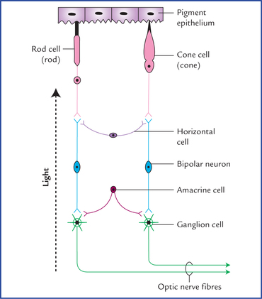
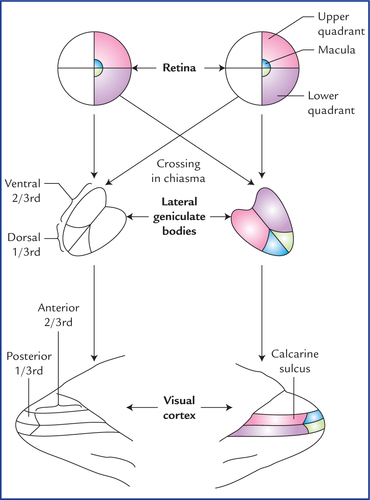
18 There are five types of special senses, viz. (a) sense of smell, (b) sense of vision, (c) sense of sound/ hearing, (d) sense of balance, and (e) sense of taste. The special senses have highly specialized receptors which provide specific information about the environment, i.e. they respond to only one type of stimulus. These receptors are located close to the brain and are well protected within the skull. Their responses are more complex but well coordinated within the brain. The special senses: (a) of smell and taste depend on chemoreceptors, (b) of vision on photoreceptors, and (c) of sound and balance on mechanoceptors. The olfactory epithelium is a specialised nasal epithelium which lines the superior one-third of the nasal cavity including the roof (Fig. 18.1). It consists of three types of cells: (a) olfactory receptor cells, (b) supporting cells, and (c) progenitor/basal cells (Fig. 18.2). Fig. 18.2 Cellular components of olfactory epithelium and the olfactory bulb. (S = stellate cell, P = periglomerular cell, OG = olfactory glands (of Bowman).) Fig. 18.4 Structures on the inferior aspect of the brain in the area surrounding the optic nerves, chiasma, optic tracts, and interpeduncular fossa. Many of these structures are related to olfactory and limbic system. The cortical zone contains olfactory glomeruli and nerve cells which form most of the prominent cellular component of the olfactory bulb. The nerve cells of the olfactory bulb comprise (Fig. 18.2): The medullary zone consists of nerve fibres of the olfactory tract. A small group of nerve cells situated at the transitional zone between the olfactory bulb and olfactory tract constitute the anterior olfactory nucleus. Olfactory bulb continues posteriorly as olfactory tract (Figs. 18.3 and 18.4). When traced posteriorly the olfactory tract divides into medial and lateral olfactory striae. The point of bifurcation is expanded and forms the olfactory trigone. The medial and lateral olfactory striae are intimately related to the anterior perforated substance and form its anteromedial and anterolateral boundaries respectively. An intermediate stria is sometimes present. It extends from the centre of trigone to anterior perforated substance where it sinks into the base of olfactory tubercle, which is a small elevation of anterior perforated substance immediately caudal to the olfactory trigone. The most of the axons of mitral cells form the lateral olfactory stria and run to the primary olfactory cortex on the same side which is located between the anterior perforated substance and the uncus on the inferomedial surface of the temporal lobe. The details are shown in Figure 18.4. The others run via intermediate olfactory stria to connect with the olfactory tubercle, and hence with the limbic system. The retina forms the inner photosensitive coat of the eyeball. It consists of two layers, an inner neural layer and an outer layer of pigment epithelium (Fig. 18.6). Fig. 18.6 Three basic layers of retina and their constituent cells. The arrow (on the left side) indicates the direction of light falling on the retina. It is important to note that several rods and cones converge on a single bipolar neuron and several bipolar neurons activate one ganglion cell. The one-to-one relationship between rods and cones, bipolar neurons and ganglion cells shown in this figure is only for the sake of simplicity. The neural layer contains three basic layers of cells: The other cells are association neurons and neuroglial cells. A small depression in the centre of the macula lutea is called fovea centralis. The fovea is about 1.5 mm in diameter and is separated from the edge of optic disc by a distance of 3 mm. The visual acuity is maximum at the fovea (i.e. clearest vision). The fovea is believed to contain only cone receptors. When one looks straight ahead with eyes fixed, that part of external world which can be seen with each eye is called visual field of that eye. Thus it is the area within which an object can be seen while the eye fixes on a spot of light or object. Laterally it extends up to 104 degree and on nasal side 65 degree. In front there is a cone-shaped area in which the visual fields of two eyes overlap. Therefore, area seen by one eye and that seen by both the eyes is more or less same except a small area that can be seen only by the eye of that side (Fig. 18.7). For the sake of convenience of description, the visual field is conventionally divided into right and left halves. Each half is further divided into an upper and a lower half, so that visual field is described to consist of four quadrants (Fig. 18.8). Fig. 18.8 Scheme to show the projection of retina on the lateral geniculate body and the visual cortex. The details areshown only for the right halves of the two retinae. In a similar manner the retina is also divided into four quadrants (Fig. 18.8). First each retina is divided into nasal and temporal halves by a vertical line passing through the fovea centralis. Then a horizontal line also passing through the fovea, divides each half of retina into upper and lower quadrants. The macular area (responsible for most acute vision) is represented separately from the peripheral parts of the retina. Light rays can enter the eye only through the pupil and since they travel in straight lines, it is obvious that objects of temporal field of vision are perceived by the nasal half of the retina whereas those in the nasal half are perceived by the temporal half of the retina.
Special Senses and their Neural Pathways
Olfactory System
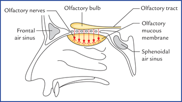
Olfactory Epithelium and Olfactory Nerves

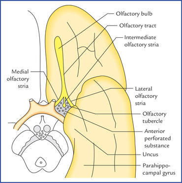
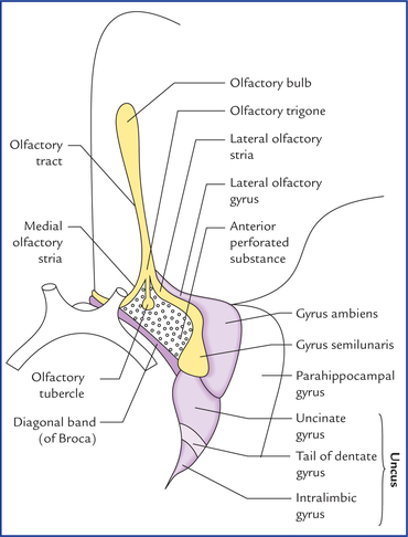
Olfactory Bulb, Tract and Striae (Figs 18.3–18.5)
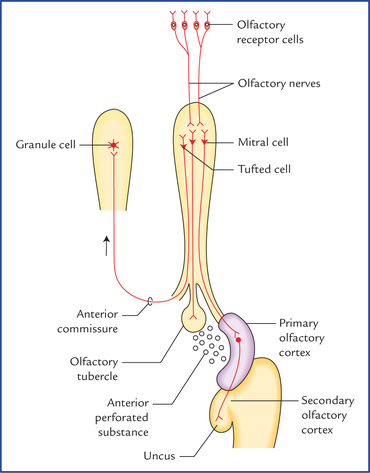
Neural Pathways for Sense of Olfaction (Fig. 18.5)
Visual System
Retina
Neural layer
Ganglion cells
Pigment epithelium
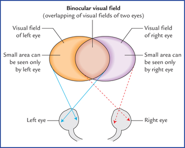
The visual field and retinal quadrants
![]()
Stay updated, free articles. Join our Telegram channel

Full access? Get Clinical Tree


Special senses and their neural pathways