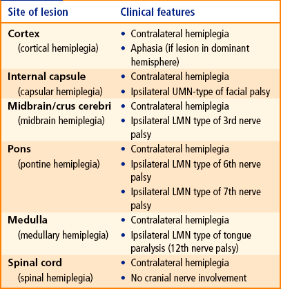
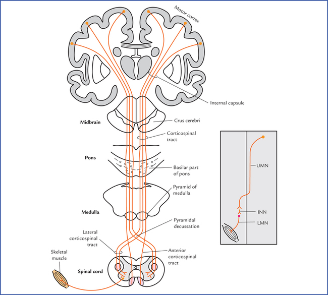
17 The somatic motor pathways of the brain and spinal cord are divided into pyramidal and extrapyramidal systems. Both these systems control the motor activities of body through lower motor neurons. The pyramidal system has a direct route to the lower motor neurons, while the extrapyramidal system has an indirect, tortuous route to these neurons. The lesions of somatic motor pathways lead to paralysis. Fig. 17.1 Course and termination of the corticospinal and corticonuclear tracts. The inset on the right side shows an abbreviated form of motor pathway. (UMN = upper motor neuron, INN = internuncial neuron, LMN = lower motor neuron.) The corticospinal tract forms a pathway that confers the speed and agility to the voluntary movements by contraction of individual or small group of muscles, particularly those moving the hands, fingers, feet and toes. Thus the integrity of corticospinal tract is essential for performing the rapid skilled voluntary movements. • The pyramidal tract contains about one million fibres in the human. • The majority of corticospinal fibres terminate on interneurons/ internuncial neurons which in turn carry the impulses to anterior horn cells. Only 9–10% synapse directly with anterior horn cells. • Fibres of lateral corticospinal tract extend to the lowest segments of the cord, while that of anterior corticospinal tract extend only up to the midthoracic level. • The longest fibres of corticospinal tract, viz. those to lower segments of cord lie most superficially, while shortest fibres lie most medially. • The fibres of corticospinal tract in addition to motor cortex, also arise from sensory cortex (one-third from premotor area and remaining one-third from primary sensory area and superior parietal lobule). The fibres arising from sensory cortex (parietal lobe) end in nucleus gracilis, nucleus cuneatus and substantia gelatinosa. They do not control motor activity but regulate the input of sensory impulses to the brain. • The representation of the musculature of the body differs at different levels. (In the primary motor cortex the body is represented upside down, in the internal capsule the motor fibres to head lie anteriorly and those for leg lie posteriorly, in the midbrain the fibres for the face lie medially while those for leg lie laterally.) In view of the frequent involvement of the pyramidal tract in cerebrovascular accidents, the arterial supply of the areas of the brain and the spinal cord occupying this tract is listed in detail in Table 17.1. Table 17.1 Arterial supply of the different parts of brain and spinal cord containing pyramidal tract N.B. The pyramidal tract is most frequently involved in cerebrovascular accident where it passes through the internal capsule. Effects of the lesions of corticospinal tracts • Spastic paralysis, due to involvement of upper motor neurons (UMN). Normally the lower motor neurons (LMNs) are under control of UMNs. Once the UMNs are damaged, they have no control on LMNs. Consequently, LMNs become hyperactive causing hypertonia or spasticity of muscles and exaggerated tendon reflexes. – Babinski’s sign is present, i.e. great toe becomes dorsiflexed and other toes fan outward when the skin along the lateral aspect of the sole of the foot is scratched with a blunt object. – Superficial abdominal reflexes are absent, i.e. abdominal muscles fail to contract when the skin of the abdomen is scratched, – Cremasteric reflex is absent, i.e. cremasteric muscle fails to contract when the skin on the medial side of the upper part of thigh is stroked. The signs and symptoms of hemiplegia differ according to the site of lesion (Table 17.2). Phylogenetically, it is an older system than the pyramidal system. It consists of all the motor tracts of the brain and spinal cord which do not pass through the medullary pyramids. The extrapyramidal system works hand in hand with the pyramidal system to perform voluntary movements (Flowcharts 17.1 and 17.2). Flowchart 17.1 Indirect motor pathways through which the corpus striatum influences the spinal cord. Flowchart 17.2 Indirect motor pathways through which the cerebral cortex influences the spinal cord. These are generally described as extrapyramidal tracts. The extrapyramidal system includes subcortical centres such as corpus striatum, globus pallidus, tectum, red nucleus, reticular formation, vestibular nuclei and neocerebellum. The corpus striatum influences descending pathways principally by its cortical connections. The other subcorti-cal centres influence the lower motor neurons in the spinal cord directly through rubrospinal, reticulospinal, tectospinal, vestibulospinal, and olivospinal tracts (Flowchart 17.2). • Postural adjustments of the body to maintain balance. • Gross synergistic voluntary movements in group of muscles affecting proximal joints of the limbs. • Movements performed unconsciously, like swinging of arms during walking. The differences between the pyramidal and extrapyrami-dal systems are given in Table 17.3. Table 17.3 Differences between the pyramidal and extrapyramidal systems N.B. Naturally occurring lesions in man rarely, if ever involve pyramidal pathway without simultaneous involvement of extrapyramidal pathways therefore the division of motor pathways into pyramidal and extrapyramidal systems is of little or no clinical relevance.
Somatic Motor and Sensory Pathways
Somatic Motor Pathways
Pyramidal System
Corticospinal (pyramidal) tract (Fig. 17.1)
Functions of the corticospinal tract (pyramidal tract)
Corticonuclear tract (Fig. 7.16)
Points to Note
Arterial supply of areas of brain and spinal cord occupied by pyramidal tract
Parts
Arterial supply
Motor cortex
• Leg area
Anterior cerebral artery
• Face, trunk and arm areas
Middle cerebral artery arm areas
Internal capsule
Branches of middle cerebral artery
Midbrain (cms cerebri)
Posterior cerebral artery
Pons
Pontine branches of basilar artery
Medulla
Medullary branches of vertebral artery
Spinal cord
Segmental branches of anterior spinal artery
Extrapyramidal System
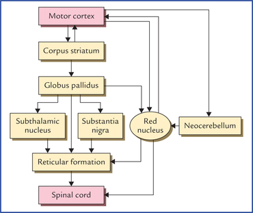
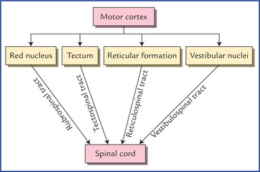
Components of extrapyramidal system
Functions of extrapyramidal system
Pyramidal system
Extrapyramidal system
Phylogeny
Phylogenetically recent in acquisition, present only in mammals and achieving its greatest development in man
Phylogenetically older than pyramidal system
Function
Responsible for non-postural, precise movements of small muscles involved in skilful activity
Responsible for gross postural (stereotyped) movements involving large groups of muscles
Pathways
Connected directly to the lower motor neurons. Therefore impulses reach the LMNs, through a direct route
Connected indirectly (polysynaptic pathway) to lower motor neurons. Therefore, impulses reach the LMNs through a circuitous route
Effects of lesion
No increase in muscle tone
Muscle tone increased (spasticity)
Cortical fibres
Arise predominantly in primary motor area (Brodmann’s area 4)
Arise predominantly in premotor area (Brodmann’s area 6)
Subcortical centres and basal ganglia
Play no role in pyramidal system
Play a key role in extrapyramidal system
![]()
Stay updated, free articles. Join our Telegram channel

Full access? Get Clinical Tree


Somatic motor and sensory pathways