111 Progressive Multifocal Leukoencephalopathy

Case 111 Progressive Multifocal Leukoencephalopathy


Ravi Pande, Maya Nader, and Remi Nader


Image

Fig. 111.1 Axial fluid-attenuated inversion-recovery magnetic resonance image of the brain.


Image 
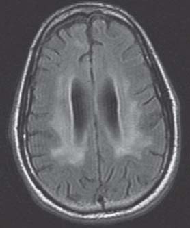
Clinical Presentation


  • A 30-year-old African American man who is deaf presented to your clinic with balance problems and frequent falls. His mother has noticed these symptoms over the past 4–6 months.
  • The symptoms are progressively getting worse; now he has to use a walker to ambulate.
  • Clinical examination reveals intact cranial nerves, except for deafness. Upper extremity strength is 5/5, lower extrem ity strength is 4+/5, and he is significantly ataxic while ambulating.

Image Questions


  1. What is your next step in the workup of this patient?
  2. Describe the magnetic resonance imaging (MRI) sfindings seen on Fig. 111.1.
  3. What is your differential diagnosis?
  4. What more of the history would you need after reviewing this MRI?

    In this case, his aunt did provide the information that he was in multiple same-sex sexual relationships.


  5. What further tests would you order?
  6. What surgical options would you consider? Under what circumstance?

    Test results obtained are as follows. The patient is human immunodeficiency virus (HIV)-positive with a viral load of 750,000 and a cluster of diff erentiation 4 (CD4) count of 61. Cerebrospinal fluid (CSF) analysis yielded protein level of 105. Polymerase chain reaction (PCR) for JC-virus was negative. CSF immunoglobulin G (IgG) index was high with nine oligoclonal bands (probable markers of demyelination and blood–brain barrier leakage).


  7. What are the yields of a brain biopsy in a patient with acquired immunodeficiency syndrome (AIDS)?
  8. Discuss your diagnosis after reviewing the test results.
  9. What is your management?
  10. What is the prognosis?

ImageAnswers


  1. What is your next step in the workup of this patient?

    • Obtain an MRI of the brain.
    • Obtain a neurology consultation.
    • May also consider obtaining MRI of the cervical and thoracic spine to rule out causes of myelopathy

  2. Describe the MRI findings seen in Fig. 111.1.

    • MRI of the brain shows extensive periventricular demyelination with some degree of atrophy. On the contrast images (not shown), there was no enhancement.

  3. What is your differential diagnosis?

    • Differential diagnosis is broad and includes the following1:

      • Multiple sclerosis (lesions should typically enhance with contrast)
      • Acute disseminated encephalomyelitis (ADEM)
      • Progressive multifocal leukoencephalopathy (PML)

    • Other less likely diagnosis include

      • Neoplasms (primary or metastatic – atypical as there is no enhancement, no mass eff ect, and lesions are diffuse)
      • Infectious causes (herpes encephalitis – atypical location)
      • Vascular (small vessel disease – also atypical location)
      • Medication or vitamin deficiencies (vitamin B6 [pyridoxine] deficiency)

  4. What more of the history would you need after reviewing this MRI?

    • Important history facts to seek include

      • Medical and surgical history
      • Medications taken
      • Family history
      • Other social history

        • Drugs, alcohol, smoking
        • Diet history
        • Sexual contacts and relationships

  5. What further tests would you order?

    • Toxicology screen
    • HIV panel
    • CSF analysis, including

      • Usual tests (cell count, cytology, cultures, glucose, protein)
      • Viral analysis – particularly PCR for JC-virus2
      • CSF immunoglobulin G index
      • Toxoplasmosis titers

  6. What surgical options would you consider? Under what circumstance?

    • A brain biopsy may be considered if the tests remain inconclusive.

      • This is particularly useful in patients with AIDS who have intracerebral lesions and negative toxoplasmosis titers.3

    • There is no focal lesion or mass effect. Therefore, there are no further indications for surgical intervention (aside from obtaining a diagnostic biopsy).4

  7. What are the yields of a brain biopsy in a patient with AIDS?

    • Overall yields range from 67–92%.3,57
    • In a study on 246 patients with AIDS, a definitive diagnosis was obtained in 92.3% of patients who underwent a biopsy.3

      • Lymphoma was the most frequent diagnosis (52.9% of patients).
      • This was followed by progressive multifocal leukoencephalopathy (18.9% of patients).
      • After lymphoma, toxoplasmosis (8.1% of patients) was the most frequently diagnosed.

    • Of note, however, is that a contrast-enhancing lesion will yield a diagnosis more frequently than a non-contrast-enhancing lesion.


      • In a study on 25 AIDS patients, the proportion of biopsies of contrast-enhancing lesions that were diagnostic and contributing to the patients’ therapeutic management was 87.5%. However, only 67% of the biopsies of nonenhancing lesions were diagnostic, and none of these lesions were treatable.5

  8. Discuss your diagnosis after reviewing the test results.

    • HIV with AIDS
    • Probable PML8

      • PML is a rare subacute demyelinating disease caused by an opportunistic papovavirus called JC-virus (JCV) or in some cases by the SV-40 strain.
      • It occurs in patients with defective cell-mediated immunity (2–5% of patients with AIDS), 9 in 1 in 1000 patients treated with Tysabri (Natalizumab; biogen idec, Cambridge, MA; immunosuppressant used in the treatment of multiple sclerosis),10 in patients with carcinoma and sarcoidosis, and in therapeutically immunosuppressed patients (e.g., transplant patients).
      • A few cases have been reported in the absence of underlying disease.
      • Pathology reveals eosinophilic intranuclear inclusions of papovavirus causing destruction of the oligodendroglia with relative sparing of axons.
      • Demyelination is most prominent in the subcortical white matter, with involvement of cerebellum, brainstem, or spinal cord white matter being less common.
      • With disease progression, the demyelinated areas coalesce to form large lesions.
      • Clinical manifestations are diverse and related to the location and number of lesions.9


        • Onset is subacute to chronic with focal or multifocal signs (hemiplegia, sensory abnormalities, visual field cuts, and other focal signs of lesion in the cerebral hemisphere).
        • Cranial nerve lesions, ataxia, and spinal cord involvement are less common.
        • Dementia ensues as the number of lesions increase.

      • A definitive diagnosis of PML can be made by brain biopsy.
      • CSF is usually normal.
      • JC virus DNA can be detected in CSF by nested PCR (n-PCR).11

  9. What is your management?

    • There is no definitive treatment available for PML.
    • Highly active antiretroviral therapy should be initiated with the goals as follows:12

      • Improvement of CD4 count
      • Normalization of viral load
      • Neurologic stabilization of PML symptoms

  10. What is the prognosis?

    • The diagnosis of PML has an overall grim outlook.11
    • The course of PML usually lasts months with 80% of patients dying within 9 months.
    • Rarely, survival may be several years, with the longest verified course being 6 years.
< div class='tao-gold-member'>

Only gold members can continue reading. Log In or Register to continue

Stay updated, free articles. Join our Telegram channel

Jul 16, 2016 | Posted by in NEUROSURGERY | Comments Off on 111 Progressive Multifocal Leukoencephalopathy

Full access? Get Clinical Tree

Get Clinical Tree app for offline access