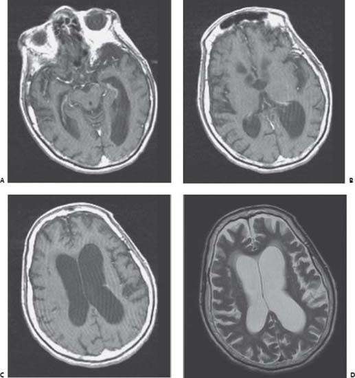
Case 113 Normal Pressure Hydrocephalus Fig. 113.1 (A–C) Sequential axial T1-weighted magnetic resonance images of the brain with infusion of intravenous contrast. (D) A T2-weighted axial image at the level of the lateral ventricles is shown.
 Clinical Presentation
 Questions
 Answers
< div class='tao-gold-member'>
113 Normal Pressure Hydrocephalus
Only gold members can continue reading. Log In or Register to continue

Full access? Get Clinical Tree


