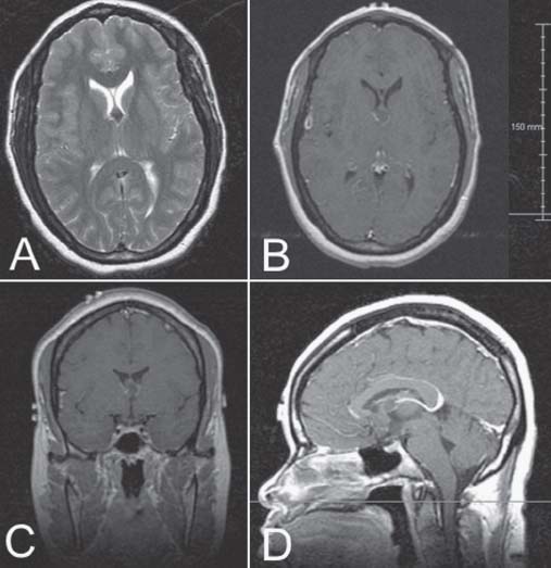
Case 26 Colloid Cyst of the Third Ventricle Fig. 26.1 (A) T2-weighted axial magnetic resonance image (MRI) at the level of the foramen of Monroe. (B) Corresponding T1-weighted MRI with contrast. (C) Coronal and (D) midsagittal MRI with contrast.
 Clinical Presentation
 Questions
 Answers
 in front, and ‧ behind the coronal suture
Complications include3,6,7
26 Colloid Cyst of the Third Ventricle
Only gold members can continue reading. Log In or Register to continue
Tags: Neurosurgery Case Review
Jul 16, 2016 | Posted by admin in NEUROSURGERY | Comments Off on 26 Colloid Cyst of the Third Ventricle

Full access? Get Clinical Tree


