67 Pediatric Head Trauma

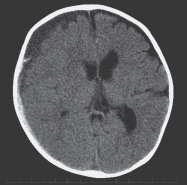
Case 67 Pediatric Head Trauma


Jeffrey Atkinson, José Luis Montes, and Abdulrahman J. Sabbagh


Image Clinical Presentation


Image

Fig. 67.1 Computed tomography scan of the brain revealing extraaxial fluid collection along the right convexity.


Image Questions


  1. What are the findings on the CT scan?
  2. What is the differential diagnosis?
  3. What other points on history and physical examination might be important?
  4. What other investigations and consultations are relevant?
  5. What is the acute management of the patient?

    The patient was taken urgently to the operating room for decompression and evacuation of the subdural hematoma. He recovered well from the injury.


  6. What are the principles of intracranial pressure (ICP) management in a child?
  7. What is the epidemiology of inflicted trauma?
  8. Is there a constellation of clinical and neurologic findings that are often associated with nonaccidental trauma?
  9. Are retinal hemorrhages pathognomic of nonaccidental trauma?
  10. What is the differential diagnosis with the association of retinal hemorrhage and bilateral subdural hematomas?
  11. What are the most common findings associated with nontraumatic head injury on CT scan?
  12. What are the mechanisms of head injury involved in infants and young children?
  13. What is the prognosis in a child with an abusive head injury?
  14. What are the medicolegal implications of inflicted trauma?

Image Answers


  1. What are the findings on CT scan?

    • The CT scan of the head demonstrates subdural collections over the right hemisphere, which are both acute and chronic.
    • There is associated mass effect on the adjacent hemisphere with compression of the ipsilateral ventricle, loss of sulcations, and midline shift.

  2. What is the differential diagnosis?

    • This subdural hematoma (SDH) is most probably traumatic.
    • The acute and chronic components may indicate hemorrhages of different ages or they may indicate an acute subdural hematoma on top of an enlarged subarachnoid space such as in the syndrome of benign extraaxial fluid collection of infancy.
    • Nontraumatic causes of SDH are very rare in infants but might include vascular malformation (of which there is no evidence on these images), coagulation disorder, or inborn error of metabolism.

  3. What other points on history and physical examination might be important?

  4. What other investigations and consultations are relevant?

    • Consultations should be obtained from the local child abuse investigative team, which might include pediatrics and social work.1
    • Ophthalmology should be consulted to examine the fundi properly to look for retinal hemorrhages that might further suggest abusive injury.4
    • Skeletal survey or nuclear medicine bone scan should be done to look for fractures.4
    • Magnetic resonance imaging (MRI) of the brain might be considered to better evaluate for cortical or parenchymal injury to the brain and perhaps to better define the ages of the subdural blood.
    • A standard investigation of abnormal coagulation would also be of benefit.1

  5. What is the acute management of the patient?

    • Acute management of this patient involves airway protection as already established by intubation.
    • ICP control medically can be attempted by mannitol, controlled ventilation, sedation of the child, which would imply the insertion of an ICP monitor.5
    • Seizure control should be obtained by an intravenous load of the antiepileptic of choice, usually phenytoin or phenobarbital.
    • In this case with the mass effect, the clinical evidence of high ICP and the clinical state of the patient, the argument for evacuation of this clot can be made. This is probably best done by craniotomy, though burr hole or transfontanel evacuation of the chronic component could be considered.
    • An ICP monitor should be inserted into the patient at the time of surgery for postoperative management.

  6. What are the principles of ICP management in a child?

    • The principles of ICP management in a child are very much the same as those in an adult.
    • A child such as this patient with a GCS <8 and a positive CT scan should be monitored invasively.5
    • Even in an infant such as the one in this case, physical examination of the fontanelle is not necessarily adequate for monitoring.
    • With the clot evacuated and the monitor in place, controlled ventilation, cerebrospinal fluid (CSF) drainage if possible, sedation, and osmotic agents can be used.6
    • In children, there is also evidence of the benefit of continuous infusions of hypertonic saline in addition to mannitol.7

  7. What is the epidemiology of inflicted trauma?

  8. Is there a constellation of clinical and neurologic findings that are often associated with nonaccidental trauma?

  9. Are retinal hemorrhages pathognomic of nonaccidental trauma?

    • No, they can be associated with normal vaginal delivery.
    • Other factors associated with retinal hemorrhages include accidental trauma, coagulopathy, hypertension, subarachnoid hemorrhage, subdural hemorrhage, papilledema, arterial hypertension, and resuscitation.11

  10. What is the differential diagnosis with the association of retinal hemorrhage and bilateral subdural hematomas?

    • Accidental trauma
    • Osteogenesis imperfecta
    • Blood coagulation dyscrasias
    • Metabolic disorders such as glutaric acidemia type I11

  11. What are the most common findings associated with nontraumatic head injury on CT scan?

    • Acute subdural hematoma9
    • Interhemispheric hemorrhage, particularly posterior or layering the tentorium
    • Parenchymal hypodensities sometimes presenting as a black brain, but most common as hemispheric or patchy hypodensities

  12. What are the mechanisms of head injury involved in infants and young children?

    • The development of acute SBH with parenchymal contusions and significant symptoms is most likely a combination of tangential acceleration, usually associated with shaking episodes and impact manipulations that are caused by hitting the head of the child against a blunt surface or throwing him against one.
    • The forces associated to impact manipulation are in the order of 20 to 30 times greater than the forces generated by shaking alone; the time lapse is significantly shorter.
    • The forces necessary to develop acute subdural hematomas, brain contusion, and diffuse axonal injury are most likely related to impact manipulation or a combination of both.
    • Low-height free-fall forces are enough to cause skull fractures or nonsymptomatic subdural hemorrhages, but not acute subdural hemorrhage accompanied by significant acute neurologic deficit as observed in most nonaccidental trauma.
    • Occasionally, low-height free falls may cause epidural hematomas that are highly symptomatic, but this represents the exception.9,12,13

  13. What is the prognosis in a child with abusive head injury?

    • In general prognosis with abusive head injury is quite poor, probably due to extensive cortical and parenchymal injury of the associated brain and even cervical medullary junction.9,14
    • There is an overall 15 to 38% mortality rate. 30 to 50% of survivors will have cognitive or other neurological deficits, and ~30% will have no significant sequelae9
    • However, in this case there appears to be very little cortical damage on the CT scan. There needs to be MRI and clinical confirmation. There might be reason to be optimistic if the clinical evolution after surgery is positive.

  14. What are the medicolegal implications of inflicted trauma?

    • The neurosurgeon is often called to establish whether there is evidence of nonaccidental trauma.
    • The neurosurgeon should be in close contact with the child protection team and ensure that all tests and studies have been done to document and support the diagnosis.
    • The neurosurgeon should be forthcoming with his or her opinion after careful consideration. Unfortunately, in medicine, answers are sometimes not clear-cut and proof of child abuse may be difficult to establish.
< div class='tao-gold-member'>

Only gold members can continue reading. Log In or Register to continue

Stay updated, free articles. Join our Telegram channel

Jul 16, 2016 | Posted by in NEUROSURGERY | Comments Off on 67 Pediatric Head Trauma

Full access? Get Clinical Tree

Get Clinical Tree app for offline access