36 Middle Cerebral Artery Aneurysm

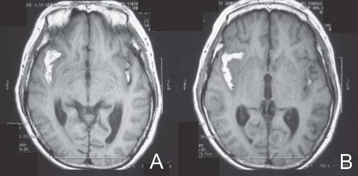
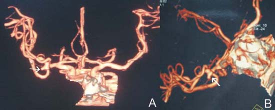
Case 36 Middle Cerebral Artery Aneurysm


Julius July and Eka Julianta Wahjoepramono


Image

Fig. 36.1 T1- weighted magnetic resonance image without contrast performed on day 4 after patient’s severe headache revealing subarachnoid blood in the sylvian fissure seen in (A) and (B).


Image

Fig. 36.2 Computed tomography angiogram with three-dimensional reconstruction confirming the presence of a right middle cerebral artery aneurysm (arrow in [A] and [B]). The aneurysm has a “Mickey Mouse” appearance and originates from the bifurcation of right M1 segment of the middle cerebral artery.


Image Clinical Presentation


Image Questions


  1. Describe your diagnosis.

    The patient obtains a computed tomography angiogram (CTA) (Fig. 36.2).


  2. What is your definitive choice of treatment? Describe the role of endovascular treatment.
  3. Describe a surgical strategy for this case.
  4. What are the late complications of middle cerebral artery (MCA) aneurysm rupture?
  5. What are the rates of morbidity and mortality based on treatment modality? What would these rates be if this was an unruptured aneurysm?

Image Answers


  1. Describe your diagnosis.

  2. What is your treatment plan? Describe the role of endovascular treatment.

    • Aneurysms arising from the MCA almost exclusively occur at the primary bifurcation and point away from the axis of the middle cerebral artery trunk. Less commonly, MCA aneurysms may arise at the origin of the anterior temporal artery, lenticulo-striate artery, secondary bifurcation, or even more distally.
    • Direct microsurgical clipping is the most common method used to treat MCA aneurysm. Even in institutions in which endovascular coiling is the preferred treatment modality, the overwhelming majority of MCA aneurysms are still treated with microsurgical clipping. MCA aneurysms are less suitable for endovascular coil packing because of both their anatomy and their frequent association with expanding hematomas. Typically, these aneurysms have wide necks with major arterial branches arising at the aneurysm base, making surgical clipping the most eff ective treatment. Because of their peripheral location and relatively straightforward surgical anatomy, microsurgical clipping of these aneurysms is easier than in other locations.2

  3. Describe a surgical strategy for this case.

    • A right pterional craniotomy will be best suitable for this case.
    • The following are some important surgical pointers pertinent to this case:

      • Operative planning should consist of patient head positioning, extent of bony exposure, necessity of placement of a ventriculostomy to relax the brain via drainage of cerebrospinal fluid (CSF), and planning for evacuation of hematoma.
      • The patient’s head is turned to the left side, around 30–45 degrees toward the floor.
      • Sharp surgical dissection of the sylvian fissure is done to identify the parent vessels, distal vessels, and the aneurysm.
      • Avoid blunt dissection around the aneurysm, as this can precipitate an intraoperative rupture. If necessary, it is better and safer to sacrifice a thin area of cortex around the aneurysm to get more working space.
      • Use of a temporary clip may be necessary, as such proximal exposure may be needed (internal carotid artery [ICA], proximal MCA). Always begin with gentle sylvian fissure dissection; the aim is to identify the parent artery for proximal control if necessary, and of course to get the aneurysm.
      • Excessive brain retraction leads to postoperative brain swelling and unsatisfactory results, regardless of how carefully the aneurysm clips are applied, and can be a significant cause of morbidity.
      • Choose the appropriate clip.2,3

  4. What are the late complications of MCA aneurysm rupture?

    • Despite good technical results, patients with ruptured MCA aneurysms often have surprisingly poor outcomes. This is primarily due to the aneurysm rupture itself, which often produces both subarachnoid and intracerebral blood causing significant morbidity.
    • Late complications of MCA aneurysm rupture include

      • Seizure: The incidence varies between 7–25% with one MCA aneurysm. It can reach 27% in multiple aneurysm cases. Most MCA aneurysms that present with temporal intracerebral hematoma (ICH) develop delayed seizures.
      • Weakness with or without dysphasia: More proximal aneurysms are associated with more severe neurologic deficits.
      • Visual-field deficit: Associated with lesions in the loop of Meyer as part of the visual pathways4,5

  5. What are the rates of morbidity and mortality based on treatment modality? What would these rates be if this was an unruptured aneurysm?

    • Overall surgical morbidity rates are ~30% for ruptured aneurysms.6
    • See Table 36.1 for more details.6,7
< div class='tao-gold-member'>

Only gold members can continue reading. Log In or Register to continue

Stay updated, free articles. Join our Telegram channel

Jul 16, 2016 | Posted by in NEUROSURGERY | Comments Off on 36 Middle Cerebral Artery Aneurysm

Full access? Get Clinical Tree

Get Clinical Tree app for offline access