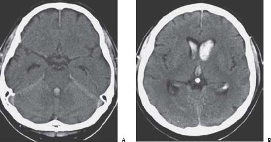
38 Intraventricular Hemorrhage

Case 38 Intraventricular Hemorrhage


Hosam Al-Jehani, Denis J. Sirhan, and Abdulrahman J. Sabbagh


Image

Fig. 38.1 Computed tomography scan without contrast at the level of (A) the 4 th ventricle and (B) foramen of Monro revealing extensive intraventricular hemorrhage with mild ventricular dilatation.


Image Clinical Presentation


Image Questions


  1. How would you classify intraventricular hemorrhage (IVH)?
  2. What are the pathophysiologic consequences of IVH?
  3. Could the clinical picture help differentiate between primary and secondary IVH?
  4. How would you grade IVH?
  5. How would you grade hydrocephalus associated with IVH?
  6. What are the treatment options for IVH?
  7. What is the prognosis of IVH?

Image Answers


  1. How would you classify IVH?

    • Intraventricular hemorrhage is classified into1,2

      • Primary IVH: in which the bleeding occurs directly into the ventricle, from a source or lesion that is in contact with or is part of the ventricular wall. This represents ~3% of all spontaneous intracerebral bleeding (intracerebral hemorrhage; ICH).
      • Secondary IVH: More common, in which the blood is introduced into the ventricular system either by dissection of an ICH or spill out of subarachnoid hemorrhage (SAH).
      • For causes of IVH see Table 38.1.2

  2. What are the pathophysiologic consequences of IVH?

    • The cerebrospinal fluid (CSF) has limited fibrinolytic activity that renders it unable to dissolve the blood clots at critical areas of the ventricular system.
    • This will result in acute obstructive hydrocephalus that could be associated with morbidity or even mortality if the intracranial pressure (ICP) is not controlled.
    • The concept is further supported by the observation that IVH volume may be associated with a commensurate decrease in global cerebral blood flow.1,2
    • There is a hypothesis that the enhanced morbidity associated with IVH is attributable, at least in part, to the pressure exerted by the clot on periventricular structures. This emphasizes the possible impact of a treatment that prevents IVH or limits further IVH expansion.1
    • Blood degradation occurs slowly by hemolysis of erythrocytes and phagocytosis by macrophages.3 These events lead to an increased risk of communicating hydrocephalus secondary to the blockage of the arachnoid granulations, and carry a small risk for cerebral vasospasm.3
    • Another interesting finding with patients who present with IVH, especially those with clinical signs of high ICP, is electrocardiographic (ECG) changes in the form of focal or generalized ST-T wave changes that could be misinterpreted as an acute myocardial infarction. This phenomenon is associated with sympathetic overdrive associated with the IVH and should be self-resolving once the ICP is under control.

  3. Could the clinical picture help differentiate between primary and secondary IVH?

    • It is possible in certain circumstances to differentiate between the two types of IVH.
    • Clinical features of each type are summarized in Table 38.2.2

  4. How would you grade IVH?

    • There are two grading scales.1,4,5
    • Graeb et al.3 devised an IVH scale (maximum score is 12).

      • Lateral ventricles score

        1. Trace amount of blood or mild bleeding
        2. Less than half of the ventricle filled with blood
        3. More than half of the ventricle filled with blood
        4. Ventricle expanded and filled with blood

      • 3rd and 4th ventricles score

        1. Blood present without dilatation
        2. Ventricle expanded and filled with blood

      • Each ventricle is scored separately, including both lateral ventricles.

    • LeRoux et al.6 described another IVH scale (Maximum score is 16).

      • Lateral, 3rd, and 4th ventricles score

        1. Trace of blood
        2. Less than half of the ventricle filled with blood
        3. More than half of the ventricle filled with blood
        4. Ventricle expanded and filled with blood

      • Each ventricle is scored separately, including both lateral ventricles.

  5. How would you grade hydrocephalus associated with IVH?

    • Diringer et al.7 described a hydrocephalus scale (Maximum score is 24.).
    • See Table 38.3 for details of the scale.8

  6. What are the treatment options for IVH?

    • The underlying cause must be treated.8

      • Needless to say, the primary cause of the IVH should be treated to prevent expansion or recurrence of the IVH. The blood pressure should be kept under control as appropriate for each patient, and correction of the coagulation abnormalities should be of primary importance.
      • There is no evidence that open surgical procedure will alter the natural history. On the contrary, they have been reported to portray worse prognosis. Some authors advocate the use of neuroendoscopy for the breakage and clearance of IVH followed by the insertion of unilateral or bilateral external ventricular drain (EVD) for further drainage.9

    • EVD5

      • Aids in relieving the hydrocephalus, if present, and helps the clearance of the CSF. This could be associated with the risk of catheter blockage that could necessitate replacement if irrigation and flushing of the catheter was not successful.
      • Prolonged use could be necessary in certain cases, which could increase the rate of infection. One should keep a close surveillance on CSF cell counts and cultures to detect early signs of infection and replace the drain accordingly.
      • Bilateral catheter placement might become indicated in certain 3rd ventricular clots that blocks the outflow through the foramen of Monro sequestering the lateral ventricles.

    • Intraventricular fibrinolytic treatment5,10,11

      • It has been found in animal models as well as in human studies that intraventricular thrombolysis expedites clot resolution and reestablishment of CSF flow. This is currently achieved using recombinant tissue plasminogen activator (rt-PA). This could achieve clot clearance in as fast as 3 or 4 days. It is also shown to reduce the 30-day mortality and reduces the cohort of poor outcome state (from 67% to 32%).
      • Contraindications to thrombolysis include large intraparenchymal hemorrhage (IPH) or IPH associated with deep or prominent cerebral tissue destruction. Another contraindication is a concomitant diagnosis associated with poor prognosis or evidence of brainstem dysfunction not in keeping with intracranial hypertension.
      • A practical approach to thrombolysis mandates a CT scan postinsertion of the EVD to ensure safe placement (all the holes of the catheter need to be within the ventricle). The dose of the rt-PA is 2–4 mg (1 mg/mL solution). It is injected slowly with frequent checking of the ICP. After the dose is delivered completely, the EVD should be closed for 1 hour, provided that the ICP is controlled, after which the EVD should be opened to a loop of 2 cm above the ear to allow drainage of the CSF. Some advocate alternating closure and opening of the EVD every hour for 24 hours to avoid collapse of the ventricle and delay the clearance of the thrombolytic agent.

  7. What is the prognosis for IVH?

    • The prognosis is not necessarily poor. Poor outcome predictors include1,2,12

      • Secondary IVH associated with a large ICH or SAH
      • Anticoagulant treatment
      • Poor neurologic condition on presentation
      • Note that quantity of blood in the ventricles and presence of hydrocephalus do not necessarily correlate with outcome.12

    • Recent series reveal the following overall outcomes for IVH2,4,12:

      • Good outcome: Ã36–54%
      • Mild to moderate disability: 39–50%
      • Severe disability: 17–35%
      • Death: 8–25%
< div class='tao-gold-member'>

Only gold members can continue reading. Log In or Register to continue

Stay updated, free articles. Join our Telegram channel

Jul 16, 2016 | Posted by in NEUROSURGERY | Comments Off on 38 Intraventricular Hemorrhage

Full access? Get Clinical Tree

Get Clinical Tree app for offline access