40 Moyamoya Disease

Case 40 Moyamoya Disease


Abdulrahman J. Sabbagh, Jean-Pierre Farmer, Jie Ma, Ahmad I. Lary, and José Luis Montes


Image

Fig. 40.1 Cerebral angiogram, internal carotid artery injection, with anteroposterior (A) and lateral (B) views.


Image 
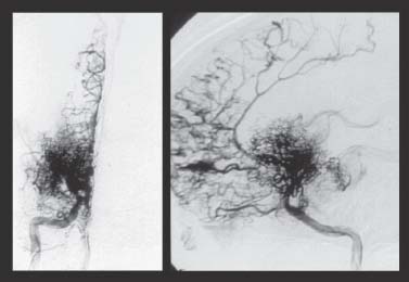
Clinical Presentation


Image Questions


  1. Describe the pertinent findings on the angiogram (Fig. 40.1).
    A physician starts her on aspirin 81 mg per day; by the time she sees you, she develops a new left foot drop.
  2. What would be your management strategy?
  3. What is the definition of moyamoya disease (MMD)?
  4. What are the clinical presentations in children for MMD?
  5. What are the factors associated with MMD?
  6. What are the angiographic stages of MMD?
  7. What is the pathophysiology of moyamoya vessel occlusion and formation?
  8. What is the natural history of MMD in the pediatric population?
  9. What are examples of revascularization procedures?
  10. Describe briefly the principles of an encephaloduroarteriosynangiosis (EDAS) procedure.

Image Answers


  1. Describe the pertinent findings on the angiogram (Fig. 40.1).

    • Left internal carotid angiography shows

      • Occlusion of the right internal carotid artery at the anterior cerebral artery (ACA)–middle cerebral artery (MCA) bifurcation
      • Extensive hypertrophy of moyamoya vessels

  2. A physician starts her on aspirin 81 mg per day; by the time she sees you, she develops a new left foot drop. What would be your management strategy?

    • Management of pediatric ischemic attack in a patient with MMD1,2:

      • Admit her to the pediatric intensive care unit.

        • Oxygenation
        • Hydration
        • Monitoring with frequent neuro checks

      • Consult pediatric neurology service to manage the new stroke.
      • Anticoagulation instead of aspirin in preparation for surgery:

        • Low-molecular-weight heparin: for example, Lovenox (Sonafi-Aventis, Bridgewater, NJ) at 0.5 mg/kg twice a day.

      • Obtain imaging studies: Computed tomography (CT) scan of the head, CT angiography or digital angiography, MRI, CT perfusion, positron emission tomography (PET) scan, etc.
      • These studies are obtained as a baseline assessment of vascular occlusion to assess extent of cerebral infarction, for documentation of new changes, and for surgical planning.
      • Preoperative planning and investigation for a revascularization procedure

  3. What is the definition of MMD?

    • Moyamoya is a Japanese term that was first used by Suzuki and Takaku in 1969.3
    • It is a radiologic term that translates as something hazy or ill-defined, like a cloud of smoke.2
    • It is a form of progressive cerebral arteriopathy characterized by2

      • Progressive occlusion of

        • Terminal internal carotid artery (ICA)
        • ACAs or MCAs

      • Profuse lenticulostriate collateral formation (moyamoya vessels) at the base of the brain

  4. What are the clinical presentations in children for MMD?

    • Clinical presentation in children includes1,4
    • Ischemic symptoms in 80%

      • Cerebral infarction
      • Transient ischemic attack

    • Epilepsy in 5%
    • Intracranial hemorrhage in 2.5%
    • Other symptoms in 12.5% (headache, movement disorders, or a mixture of symptoms)

  5. What are the factors associated with MMD?

    • Associated factors include46

      • Asian ethnicity
      • Neurofibromatosis type I, especially if with radiation for an optic nerve glioma
      • Radiation for optic or hypothalamic pathway glioma or craniopharyngioma
      • Down syndrome
      • Renal artery stenosis
      • Hypertension
      • Thalassemia and sickle cell anemia
      • Other conditions

  6. What are the angiographic stages of MMD?

    • MMD has six angiographic stages (Fig. 40.2).3,7

      • Stage 1: Narrowing of the carotid bifurcation
      • Stage 2: Initiation of the moyamoya vessels and dilatation of the intracerebral main arteries
      • Stage 3: Intensification of the moyamoya vessels and nonfilling of the ACAs and MCAs
      • Stage 4: Minimization of the moyamoya vessels and disappearance of the posterior cerebral artery
      • Stage 5: Reduction of the moyamoya vessels. The main arteries arising from the ICA disappear.
      • Stage 6: Complete loss of the moyamoya vessels. Only the collateral circulation from the external carotid artery is seen at this stage.

  7. What is the pathophysiology of moyamoya vessel occlusion and formation?

    • Pathophysiology of arterial occlusion and formation in MMD are described below (Fig. 40.3).810

      • Angiogenic factors such as CRABP-1 (cellular retinoic acid binding protein), bFGF (fibroblast growth factor), TGF-β (transforming growth factor-β), HGF (hepatocyte growth factor), and others contribute to

        • A slow and long-term proliferation and migration of intimal smooth muscle deposition eventually causing vessel occlusion
        • Revascularization and formation of the moyamoya vessels
        • Vasodilatation of collaterals

      • Other genetic and environmental factors come into play.810

  8. What is the natural history of MMD in the pediatric population?

    • The natural history of MMD in the pediatric population11

      • In patients who are not surgically revascularized, only around 23% subsequently improve, 20% do not change, and up to 57% get worse (these results are based on a study by Choi et al.11 of 52 subjects with a mean follow-up of 67.2 months).
      • 65% of patients of the same age group that are revascularized improve, 23% remain unchanged, and only 12% get worse (These results are based on a study by Choi et al.11 of 36 subjects with a mean follow-up of 28.8 months).

  9. What are examples of revascularization procedures?

    • Indirect procedures include5,11,12

      • EDAS: encephaloduroarteriosynangiosis
      • EMS: encephalomyosynangiosis
      • EDAMS: encephaloduroarteriomyosynangiosis
      • Modified EDAS (pial synangiosis)
      • EGS: encephalogaleosynangiosis
      • Omental transfer

    • Direct procedures include12

      • Superficial temporal artery to middle cerebral artery (STA–MCA) bypass

  10. Describe briefly the principles of an EDAS procedure.

    • The steps of an EDAS procedure include2 (Fig. 40.4)

      • After preparing the scalp, the STAs and occipital arteries (OcAs) are outlined using a Doppler ultrasound; then the skin overlying them is opened and the vessels identified.
      • The STAs and OcAs are separated from neighboring tissues along with a strip of adventitia on either side of the vessels.
      • A craniotomy or a small craniectomy is performed, and then a Z-shape durotomy is tailored.
      • The adventitia is sutured to close the durotomy edges, making the STAs and the OcAs in close proximity to pial vessels.
      • This facilitates collateral development over time, from the external to the intracranial circulation.
< div class='tao-gold-member'>

Only gold members can continue reading. Log In or Register to continue

Stay updated, free articles. Join our Telegram channel

Jul 16, 2016 | Posted by in NEUROSURGERY | Comments Off on 40 Moyamoya Disease

Full access? Get Clinical Tree

Get Clinical Tree app for offline access