44 High-grade Carotid Stenosis and Intracranial Aneurysm

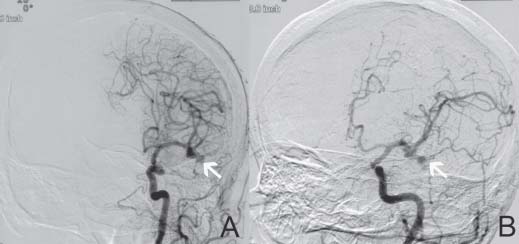
Case 44 High-grade Carotid Stenosis and Intracranial Aneurysm


Glenn C. Hunter and Remi Nader


Image

Fig. 44.1 Cerebral angiogram, left carotid injection, showing on (A) anteroposterior and (B) lateral views showing a left middle carotid artery trifurcation aneurysm of 7 mm in size.


Image Clinical Presentation


  • A 74-year-old woman presents with a history of left hemispheric transient ischemic attacks (TIAs).
  • She experienced one episode of right arm and leg weakness that completely resolved within 5–7 minutes ~4 weeks ago.
  • Her blood pressure was 160/110 mm Hg while still on two antihypertensive medications. The blood pressure in her left arm was 30 mm Hg lower than that on the right side.
  • Bilateral carotid bruits are present.

Image Questions


  1. What initial studies would you order?
  2. What imaging studies would assist in the diagnosis?
  3. How would you manage this patient initially?

    She returns 2 weeks later with complaints of having discontinued her clopidogrel (Plavix ; Bristol-Myers Squibb, New York, NY) because of bruising. She has experienced two episodes of TIAs since discontinuing clopidogrel.


  4. How do you manage her now?
  5. A cerebral angiogram is obtained. Is this preferable to computed tomography (CT) or magnetic resonance angiography (MRA)?

    The angiogram demonstrated an irregular high-grade (90%) left internal carotid artery (LICA) stenosis (not shown here) and a left 7 mm middle cerebral artery cerebral aneurysm (Fig. 44.1).


  6. What are the prevalence and sex differences of an unruptured intracranial aneurysm (UIA)?
  7. What is the incidence of a UIA on cerebral angiograms performed for carotid disease?
  8. Are there any conditions associated with intracranial aneurysms?
  9. How may intracranial aneurysms present?
  10. What are the factors that predispose to aneurysm rupture?
  11. What are the rupture rates in this case?
  12. List the therapeutic options for this patient.
  13. What factors need to be taken into consideration in selecting the treatment option?
  14. Describe the anatomical segments of the internal carotid artery and their branches.

Image Answers


  1. What initial studies would you order?

    • The patient should have a work-up for stroke.
    • Laboratory studies should include complete blood count (CBC), coagulation studies, electrolytes, and renal function tests.
    • Chest x-ray, electrocardiogram (EKG)

  2. What imaging studies would assist in the diagnosis?

    • A carotid duplex scan
    • CT or magnetic resonance imaging (MRI) of the brain

      • The duplex scan shows 80–99% LICA stenosis with 50–79% stenosis on the right side.
      • A nonenhanced CT scan shows no evidence of hemorrhage or ischemia.

  3. How would you manage this patient initially?

    • She is placed on aspirin and clopidogrel.
    • Arrangements should be made for an outpatient cerebral angiogram.

  4. How do you manage her now?

    • She is experiencing more frequent TIAs; therefore, direct hospital admission is indicated.
    • Consider intravenous heparin therapy if no hemorrhage is seen on the CT scan of the brain.

  5. A cerebral angiogram is obtained. Is this preferable to CT or MRA?

    • This patient has evidence of both subclavian and carotid stenosis. A cerebral angiogram would provide the most definitive diagnosis.
    • However, the inherent risks of this more invasive procedure (such as stroke, etc.) should be discussed with the patient prior to the study.

  6. What are the prevalence and sex differences of an UIA?

  7. What is the incidence of an UIA on cerebral angiograms performed for carotid disease?

    • Incidence: 1:40 patients with symptomatic carotid stenosis have UIAs.2

      • Based on the North American Symptomatic Carotid Endarterectomy Trial (NASCET) study,3 there is a 3.1% incidence of an UIA in cerebral angiograms performed for carotid disease.

        • 96% of aneurysms are less than or equal to 10 mm in size.
        • 55% are ipsilateral to the side of the carotid stenosis.
        • These account for 6 to 16% of subarachnoid hemorrhages (SAHs)
        • 8 to 34% are multiple.

  8. Are there any conditions associated with intracranial aneurysms?

    • UIA can have a congenital predisposition and can be associated with1

      • Aortic coarctation
      • Polycystic kidneys (autosomal dominant)
      • Family history of UIA/SAH
      • Fibromuscular dysplasia
      • Pseudoxanthoma elasticum
      • Moyamoya disease
      • Systemic lupus erythematosus
      • Arteriovenous malformations

  9. How may intracranial aneurysms present?

    • Incidental
    • SAH
    • Embolism (rare)
    • Headache
    • Cranial nerve palsy1

  10. What are the factors that predispose to aneurysm rupture?

    • Size
    • Location (anterior vs. posterior circulation)
    • Previous SAH (increases hemorrhage rate by 11-fold)4

  11. What are the rupture rates in this case?

    • Risk of rupture in this case is less than 1%. Rupture risk depends on size and location of the aneurysm.1,46
    • See Case 31 for a detailed description of the associated risks.

  12. List the therapeutic options for this patient.

    • Carotid endarterectomy (CEA) without UIA clipping (UIA observation)
    • Combined CEA and UIA clipping
    • CEA and aneurysm coiling
    • Carotid artery stenting (CAS) and aneurysm coiling7

  13. What factors need to be taken into consideration in selecting the treatment option?

    • Determine if the symptoms are of hemodynamic or embolic origin.
    • Determine which lesion is causing the symptoms.
    • Determine the relationship between the two lesions:

      • Embolic symptoms are most often due to carotid bifurcation plaque.
      • Carotid lesions are a more common cause of symptoms (as in this case).

    • Certain factors with the treatment of one lesion may exacerbate the risks of the other:

      • CEA may increase the risk of aneurysm rupture due to an increase in BP (this is very unlikely, however).8
      • Hypotension necessary to clip an aneurysm may cause thrombosis of a high-grade carotid stenosis.8

    • Based on the NASCET study,3 the 5-year risk of rupture of UIA differs based on the treatment modality of the underlying carotid disease.

      • With CEA, it is 10%.
      • With best medical therapy, it is 22.7%.

  14. Describe the anatomical segments of the internal carotid artery and their branches.
< div class='tao-gold-member'>

Only gold members can continue reading. Log In or Register to continue

Stay updated, free articles. Join our Telegram channel

Jul 16, 2016 | Posted by in NEUROSURGERY | Comments Off on 44 High-grade Carotid Stenosis and Intracranial Aneurysm

Full access? Get Clinical Tree

Get Clinical Tree app for offline access