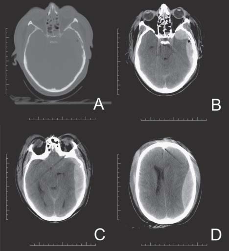
47 Epidural Hematoma

Case 47 Epidural Hematoma


Abdulrazag Ajlan and Judith Marcoux


Image

Fig. 47.1 Computed tomography scan of the head, bone window is displayed in (A) where a left temporal bone frac ture is seen. (B), (C) and (D) are brain windows showing a large epidural hematoma with significant mass effect and uncal herniation with brainstem compression and displacement (C) and significant midline shift (D).


Image Clinical Presentation


  • A 42-year-old woman fell from the third floor of a building. She presents with a Glasgow Coma Score (GCS) of 7.
  • The patient is hemodynamically stable and she is quickly intubated.
  • Her pupils are both reactive, but asymmetrical.

Image Questions


  1. Describe what you see on the computed tomography (CT) scan (Fig. 47.1).
  2. What is the pathophysiology of this intracranial bleed?
  3. What is your initial management?
  4. What are the criteria for surgical evacuation?
  5. What is the prognosis?

Image Answers


  1. Describe what you see on the CT scan (Fig. 47.1).

    • There is a linear nondisplaced temporal bone fracture with underlying pneumocephalus.
    • There are also hyperdense lesions in the temporal and temporoparietal areas on the same side of the fracture. The lesions are biconvex (lens shape) and are compatible with an epidural hematoma (EDH).
    • The lesions are causing a midline shift of 0.7 cm and a right-side uncal herniation.

  2. What is the pathophysiology of this intracranial bleed?

    • Epidural hematomas are usually located in the temporoparietal areas; posterior fossa hematomas represent 5% of the EDH.
    • The source is usually a meningeal artery, which is most commonly the middle meningeal artery, but sometimes this can be a bleeding vein or an underlying sinus.
    • The bleeding in the epidural space will strip the dura from the skull causing mass effect and raising intracranial pressure.
    • Approximately 85% of cases will be associated with skull fracture.
    • 20% of patients will present in a comatose state.1 Other presentations include localized neurologic findings and confusion. The classical presentation of transient improvement followed by sudden deterioration occurs in 47% of admissions, which is called the Lucid Interval.2

  3. What is your initial management?

    • Once the airway is secured and the patient is hemodynamically stable, she needs to undergo emergency evacuation for the hematoma.
    • The patient should also be loaded with an antiepileptic drug.
    • Infusing mannitol and hyperventilation may be done as temporary measures before the evacuation.
    • This is a surgical emergency because there is a symptomatically significant mass larger than 1 cm with midline shift greater than 0.5 cm.

  4. What are the criteria for surgical evacuation?

    • An EDH more than 30 cm3 regardless of the GCS.2
    • A patient with a GSC of more than 8 and a hematoma less than 30 cm3, less than 15 mm thickness, less than 0.5 cm midline shift, and without focal deficit can be managed by close observation with serial scans and placement in a monitored neurosurgical unit.2

  5. What is the prognosis?

    • Outcome depends on many factors, which include GCS, CT scan findings, age, timing of surgery, and the presence of other lesions.
    • The overall mortality for patients who underwent surgical evacuation is 10%.2 The faster the evacuation is done the better will be the outcome because the neurologic status prior to surgery is the main determinant of outcome.
    • EDH evacuation is considered one of the most “cost effective” of all surgical procedures in terms of quality of life.3
< div class='tao-gold-member'>

Only gold members can continue reading. Log In or Register to continue

Stay updated, free articles. Join our Telegram channel

Jul 16, 2016 | Posted by in NEUROSURGERY | Comments Off on 47 Epidural Hematoma

Full access? Get Clinical Tree

Get Clinical Tree app for offline access