48 Traumatic Acute Subdural Hematoma

Case 48 Traumatic Acute Subdural Hematoma


Abdulrazag Ajlan and Judith Marcoux


Image

Fig. 48.1 Computed tomography scan of the head, brain windows showing a right-side subdural hematoma with associated midline shift greater than 5 mm (A) and brain compression (B–D).
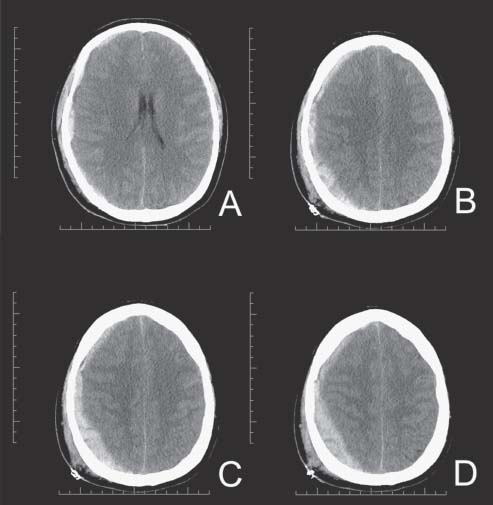

Image Clinical Presentation


  • A 39-year-old man is involved in an all-terrain vehicle accident; he had no helmet on.
  • His Glasgow Coma Scale (GCS) score on arrival in the emergency room was 7 (Eyes 1, Verbal 1, Motor 5).
  • The patient is hemodynamically stable and is quickly intubated.

Image Questions


  1. Describe what you see on the computed tomography (CT) scan (Fig. 48.1).
  2. What is the pathophysiology of this intracranial bleed?
  3. What is your management?
  4. What are the criteria for surgical evacuation for acute subdural hematomas (SDHs)?
  5. What is the prognosis?
  6. What other types of traumatic intracranial hemorrhage can you see?
  7. What are the indications for surgical evacuation in the different types of intracranial hemorrhage?
  8. What are the indications for antiepileptic medication?

Image Answers


  1. Describe what you see on the CT scan (Fig. 48.1).

    • There is an acute subdural hematoma over the right hemisphere which causes mass effect, compression of the brain, and a midline shift.

  2. What is the pathophysiology of this intracranial bleed?

    • Acute subdural hematomas are usually due to torn bridging veins at the surface of the brain. Damaged cortical arteries could also produce subdural hematomas.
    • A significant degree of impact is usually required to produce a subdural hematoma.

  3. What is your management?

    • This patient is comatose; he has a mass lesion seen on the CT scan and significant midline shift associated with it (greater than 5 mm).
    • After ensuring that there is no life-threatening injury, this patient should be taken to the operating room for quick decompression via a large frontotemporoparietal craniectomy or craniotomy.

  4. What are the criteria for surgical evacuation for acute SDHs?

    • Indications for surgery are as follows1:

      • An acute SDH with a thickness greater than 10 mm or a midline shift greater than 5 mm on CT scan should be surgically evacuated, regardless of the patient’s GCS score.
      • All patients with an acute SDH in a coma (GCS less than 9) should have an ICP monitor.
      • A patient with a GCS score less than 9 an acute SDH less than 1 mm, and a midline shift less than 5 mm should undergo surgical evacuation of the lesion if one of the following is present:

        • Decrease in the GCS score by 2 or more points
        • Asymmetric or fixed and dilated pupils
        • Elevated intracranial pressure (ICP) of more than 20 mm Hg

  5. What is the prognosis?

    • The prognosis will depend on the initial GCS score, the age, and the associated brain damage (with subsequent intracranial hypertension).
    • Early surgery may improve the outcome.

  6. What other types of traumatic intracranial hemorrhage can you see?

    • The other types of traumatic intracranial bleeding, apart from acute subdural hemorrhage and acute epidural hemorrhage, include

      • Intraventricular hemorrhage
      • Subarachnoid hemorrhage
      • Intracerebral contusions; three subtypes:

        • Coup contusion (occurs at the site of the impact)
        • Contre-coup contusion (occurs at sites remote from the impact and are due to movements of the brain inside of the skull; they are most commonly found in the frontal and temporal poles and the subfrontal area)
        • Gliding contusion (due to herniation or tissue tear)

      • Intracerebral hematomas (greater content of blood compared with contusions); usually caused by shearing of small vessels, but could also be caused by traumatic aneurysms.

  7. What are the indications for surgical evacuation in the different types of intracranial hemorrhage?

    • An intraventricular hemorrhage can cause hydrocephalus requiring cerebrospinal fluid drainage. Subarachnoid hemorrhage does not require surgical evacuation; however, if it is abundant enough, it may cause vasospasm.
    • Contusions could be small and limited to the cortical layer, but they could also cause significant mass effect. Furthermore, contusions have the tendency to increase in size over the first 24–48 hours (and sometimes even longer) following a trauma. The prognosis following traumatic contusions will depend on the location and the number and the size of the contusions. Contusions may be a cause of posttraumatic seizure. Due to their location, they can also play a role in cognitive and behavioral deficit following a traumatic brain injury (TBI).
    • Patients with mass effect, worsening neurologic exam, or refractory high ICP should undergo surgical evacuation. Patients with a GCS score of 6–8 with frontal or temporal contusions of greater than 20 cm3 with a midline shift of greater than or equal to 5 mm or compression of the basal cisterns should have surgery. Any contusion with a volume greater than 50 cm3 should be evacuated.2 Any lesion of the posterior fossa associated with mass effect or neurologic dysfunction should be evacuated.3

  8. What are the indications for antiepileptic medication?

    • Prophylaxis of late posttraumatic seizure is not recommended.
    • Prophylaxis of early posttraumatic seizure (within 7 days) is recommended, but has not been shown to change the overall outcome.4
< div class='tao-gold-member'>

Only gold members can continue reading. Log In or Register to continue

Stay updated, free articles. Join our Telegram channel

Jul 16, 2016 | Posted by in NEUROSURGERY | Comments Off on 48 Traumatic Acute Subdural Hematoma

Full access? Get Clinical Tree

Get Clinical Tree app for offline access