49 New Trends in Neurotrauma Monitoring

Case 49 New Trends in Neurotrauma Monitoring


Judith Marcoux and Abdulrazag Ajlan


Image

Fig. 49.1 Computed tomography scan of the head showing a very small intraparenchymal hematoma in the right hippocampal area.


Image 
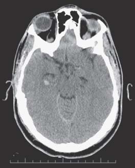
Clinical Presentation


Image Questions


  1. What other tools can help you optimize his ICP management?
  2. What is the role of continuous electroencephalography (EEG)?
  3. What is the role of microdialysis?

Image Answers


  1. What other tools can help you optimize his ICP management?

  2. What is the role of continuous EEG?

    • Posttraumatic seizures complicate a sizable percentage of TBIs. The risk will increase with the severity of the trauma.
    • In patients with moderate and severe TBI, 22.3% had electrical seizures on continuous EEG monitoring. More than half of which had no clinical manifestation of their seizures.4
    • In critically ill comatose patients, the duration of nonconvulsive status epilepticus and the delay to diagnosis was associated with increased mortality.5
    • Continuous EEG monitoring may help diagnose and treat nonconvulsive seizures, and thus prevent secondary injury to the brain. Furthermore, continuous EEG is of primary importance in the titration of barbiturate-induced coma to treat raised ICP.

  3. What is the role of microdialysis?

    • Microdialysis gives a unique insight into the brain’s metabolism by measuring tissue biochemistry.
    • It can give a warning regarding impending hypoxia/ischemia or the occurrence of secondary damage. But its measure is local and there is a great variability in the range of the values collected. Its use is still limited to research. However, a growing number of clinicians are using it. In 2004, a consensus6 was made for its use and some of the major points are as follows:

      • The use of microdialysis should be reserved for patients with a severe TBI requiring ICP monitoring.
      • The probe should be placed in the right frontal area in patients with diffuse lesions or placed in the pericontusional area, if there is a focal lesion.
      • The best marker for secondary damage is the lactate/pyruvate ratio; glucose, glutamate, and glycerol can also be used as markers of ischemia.
< div class='tao-gold-member'>

Only gold members can continue reading. Log In or Register to continue

Stay updated, free articles. Join our Telegram channel

Jul 16, 2016 | Posted by in NEUROSURGERY | Comments Off on 49 New Trends in Neurotrauma Monitoring

Full access? Get Clinical Tree

Get Clinical Tree app for offline access