50 Intracranial Pressure Management

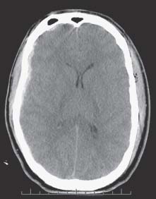
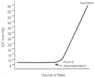
Case 50 Intracranial Pressure Management


Abdulrazag Ajlan and Judith Marcoux


Image

Fig. 50.1 A brain computed tomography scan showing a small right subdural hematoma with a midline shift of 4 mm. There is diffuse brain swelling and small ventricles.


Image

Fig. 50.2 The brain can compensate within a limited range of pressure. After the compensatory mechanisms fail, the intracranial pressure will increase dramatically.


Image Clinical Presentation


Image Questions


  1. What is the pathophysiology of raised ICP?
  2. How can we monitor the ICP?
  3. What are the indications for ICP monitoring?
  4. How can we manage elevated ICP (describe first, second, and third tier measures)?

Image Answers


  1. What is the pathophysiology of raised ICP?

    • The Monro-Kellie hypothesis states that the cranial compartment is incompressible, and the volume inside the cranium is a fixed volume.
    • The cranium and its constituents (blood, cerebrospinal fluid [CSF], and brain tissue) create a state of volume equilibrium, such that any increase in volume of one of the cranial constituents must be compensated by a decrease in volume of another.1,2
    • Small increases in brain volume does not lead to immediate high ICP because of compensatory measures such as displacement of CSF into the spinal canal, stretching of the falx cerebri and the tentorium, and decrease in venous blood volume. However, once the ICP goes beyond the compensatory phase, small increases in brain volume (or any other constituent) can lead to marked elevations in the ICP (Fig. 50.2).
    • Once the ICP starts to be elevated, it will affect the brain in two major ways:

      • High ICP will lead to decrease cerebral blood flow (CBF) as can be observed on the following equations:

        • CPP (cerebral perfusion pressure) = MAP (mean arterial pressure) – ICP
        • CBF = CPP / CVR (cerebral vascular resistance)

      • Any increase in the ICP will decrease the CPP. In the normal physiologic state, the CBF will be kept constant because the CVR will vary to compensate, this is called autoregulation. This compensation is impaired in the extreme of pressure or in abnormal states (such as brain trauma).

    • Once the ICP is high and uncompensated, the brain tissue will start shifting and herniating through dural openings. This herniation will cause brain tissue compression, disturbance in blood flow, damage to the vasculature, as well as further increase in the ICP. There are different types of herniation:

      • Uncal herniation is caused by a mass in the middle fossa that displaces the uncus between the mid-brain and tentorial edge. This causes compression on the descending tract and the reticular formation in the brainstem causing a decrease in the level of consciousness and contralateral hemiparesis. The ipsilateral oculomotor nerve is also affected causing ptosis and mydriasis (Fig. 50.3 and Fig. 50.4)
      • Sometimes the midbrain is squeezed against the contralateral tentorial edge (Kernohan’s notch) causing ipsilateral hemiparesis (Fig. 50.3 and Fig. 50.4). The posterior cerebral artery will be compressed causing ischemia
      • With more severe herniation, the basilar artery will be stretched causing tearing of the brainstem perforator vessels, brainstem infarction, and bleeding (Duret’s hemorrhages)
      • The Cushing triad occurs from high ICP and herniation. This triad includes hypertension, bradycardia, and respiratory irregularities.

  2. How can we monitor the ICP?

    • One of the most reliable methods is the clinical picture of the patient. If the patient is awake enough to be followed clinically, this can be used as an objective parameter indicating that he is maintaining his perfusion and that his ICP is compensated. Once the clinical exam is lost for any reason (such as decrease level of consciousness or intoxication), another parameter should be used.
    • Several methods have been developed for measuring the ICP. The classical methods include

      • Intraventricular catheter placement (ventriculostomy)
      • Intraparenchymal monitoring
      • Epidural devices
      • Subdural/subarachnoid devices (bolts)

    • The ventricular catheter is the gold standard given that it is the most accurate. It is also therapeutic as well as diagnostic.

  3. What are the indications for ICP monitoring?

    • The guidelines were revised by the Brain Trauma Foundation in 20073 and include

      • Patient with severe injury (GCS ≤8) after resuscitation and with an abnormal CT scan (hematoma, contusion, edema).
      • Patient with severe injury (GCS ≤8) and normal CT scan with two or more of the following:

        • Age above 40 years
        • Unilateral or bilateral motor posturing
        • Systolic blood pressure <90 mm Hg

    • ICP monitoring is not routinely indicated in mild or moderate brain trauma cases. However the treating physician can choose to monitor ICP in certain conscious patients with abnormal CT scan findings.3

  4. How can we manage elevated ICP (describe first, second, and third tier measures)?

    • Brain injury from trauma results from

      • Primary injury from the first impact. This type of damage has no treatment other than prevention
      • Secondary injury, which can be due to

        • Intracranial causes such as expanding hematomas, contusions, or diffuse edema, which will cause high ICP
        • Systemic causes such as hypotension, hypoxia, and pyrexia, will cause mismatch in the metabolic demand and the blood flow.

    • Therefore, the treatment starts at the scene by preventing hypotension and hypoxia with adequate resuscitation and airway management, followed by a rapid transfer to a trauma center.
    • Once the CT scan is done and a surgical mass lesion is ruled out, an ICP monitor is inserted if indicated.
    • The goal of treatment is to keep an adequate blood flow which will match the metabolic demand of the injured brain. To reach this goal, the ICP should be kept below 20 mm Hg,4 and the CPP between 50 and 70 mm Hg.5 First, second, and third tier therapies have been devised for ICP treatment.
    • First tier therapies:

      • Elevation of the head to 30 degrees to enhance the venous outflow
      • Keeping the neck straight and the venous outflow patent.
      • Ventilation to normocarbia (pCO2 = 35–40 mm Hg) to prevent cerebral vasodilatation. Hyperventilation can reduce ICP by causing cerebral vasoconstriction. On the other hand, this decrease in CBF can cause ischemia. Therefore, the use of hyperventilation as a prophylactic measure with pCO2 <25 mm Hg is not recommended, especially in the early period after trauma. Hyperventilation is only recommended as a temporary measure in the case of high ICP until other treatments are started or with other monitoring tools for the cerebral blood flow (see Case 49, New Trends in Neurotrauma Monitoring).
      • Avoid hyperthermia because it may lead to an increase in the metabolic rate. Acetaminophen and a cooling blanket can be used to achieve this goal.

    • Second tier therapies:

      • Agitation and pain are two common causes of elevated ICP. Thus, sedation and pain control present an effective treatment modality.
      • Hyperosmolar treatment is used to decrease brain edema and subsequently the ICP. The hyperosmolar treatment will help also in cerebral perfusion.6 Mannitol and hypertonic saline can be used as osmotic therapy agents.
      • Neuromuscular paralysis for maximal muscle relaxation and decrease muscle tone
      • CSF drainage from the intraventricular drain

    • Third tier therapies:

      • Decompressive craniectomy can be very effective in reducing the ICP. However, it is not clear if this measure improves the outcome. There are no clear guidelines about indications and timing. The only randomized clinical trial was done in a pediatric group. It shows a better outcome, but this did not reach statistical significance.7
      • Barbiturate coma: High-dose barbiturate therapy can result in control of ICP when all other medical and surgical treatments have failed. This effect is attributed mainly to the decrease in the cellular metabolic rate and subsequent decrease in CBF. The main side effects are hemodynamic instability and lowered immunity. It has shown no clear benefit in improving outcome and the prophylactic use in the treatment of ICP is not recommended.8
< div class='tao-gold-member'>

Only gold members can continue reading. Log In or Register to continue

Stay updated, free articles. Join our Telegram channel

Jul 16, 2016 | Posted by in NEUROSURGERY | Comments Off on 50 Intracranial Pressure Management

Full access? Get Clinical Tree

Get Clinical Tree app for offline access