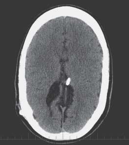
55 Slit Ventricle Syndrome

Case 55 Slit Ventricle Syndrome


Jeffrey Atkinson


Image

Fig. 55.1 Computed tomography scan of the brain revealing relatively small lateral ventricles and ventriculoperitoneal shunt located within the ventricle.


Image Clinical Presentation


Image Questions


  1. What is the differential diagnosis?
  2. What is the next possible step in the workup of this patient?

    Shunt revision is performed, and at the time of surgery the distal and proximal catheters are changed due to “sluggish” flow. She is relieved of her headaches for a few hours, but the symptoms return shortly thereafter.


  3. What are the diagnostic possibilities now?
  4. What is the next step in her management?

    CT scan remains unchanged; nuclear medicine shuntogram shows good flow.


  5. What are other potential investigations?

    Intracranial pressure (ICP) monitoring shows readings generally between 5 and 15 mm Hg depending on her position with no single reading higher than 20 mm Hg, and no reading lower than –5 mm Hg.


  6. Describe the different types of syndromes associated with headaches and stable ventricle size on CT scan and outline management strategies for each.

Image Answers


  1. What is the differential diagnosis?


    • Headache of the usual types (tension, migraine, etc.)
    • Headache related to shunt malfunction
    • Headache related to overshunting
    • It is essential to consider shunt malfunction even in the presence of unchanged or small ventricles. It may be of benefit to know the details of her last shunt revision with respect to headache history and changes seen on CT scan.

  2. What is the next possible step in the workup of this patient?


  3. What are the diagnostic possibilities now?


    • Early return of symptoms after shunt revision always prompts an appropriate workup for shunt malfunction.
    • A surgery can always convert a working shunt to a nonworking shunt. In the absence of any new signs of shunt malfunction, the same diagnostic possibilities as stated previously apply.

  4. What is the next step in her management?


    • This patient needs an early-repeat CT scan.
    • If there is still no change in her ventricles, the management could proceed according to Question #2.

  5. What are other potential investigations?


    • At this stage, ICP monitoring becomes the best option.

  6. Describe the different types of syndromes associated with headaches and stable ventricle size on CT scan and outline management strategies for each.


    • Patients with VP shunts, new headaches, and no change in ventricular size on their CT scan can be broken down into four categories (variably described as part of the “slit ventricle syndrome”):


      • Shunt is malfunctioning despite CT findings – this can be determined by a shuntogram or shunt exploration, and the treatment is shunt revision.3
      • Shunt is overdraining – ICP monitoring may be necessary, though clinical history can be helpful and upgrading of valve to a higher resistance setting would be the appropriate treatment.3,4
      • Shunt is functioning maximally, but ICP is high anyway – this is rare and can only be really established with shunt exploration and ICP monitoring. In this rare instance, cranial expansion procedures would be the treatment of choice.3
      • Not a shunt problem – established with normal ICP tracing with ICP monitor, and treatment is neurology and/or pain service consultation, and other medical management for chronic headache relief.2
< div class='tao-gold-member'>

Only gold members can continue reading. Log In or Register to continue

Stay updated, free articles. Join our Telegram channel

Jul 16, 2016 | Posted by in NEUROSURGERY | Comments Off on 55 Slit Ventricle Syndrome

Full access? Get Clinical Tree

Get Clinical Tree app for offline access