59 Brainstem Glioma 2: Medulla Oblongata

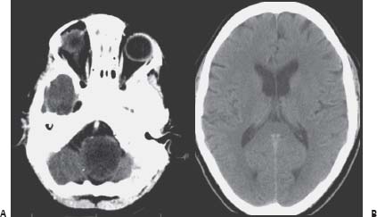
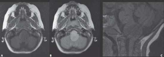
Case 59 Brainstem Glioma 2: Medulla Oblongata


Jean-Pierre Farmer, Abdulrahman J. Sabbagh, and Ahmad Al-Jishi


Image Clinical Presentation


  • A 4-year-old boy presents to the emergency room sent by his pediatrician for head tilt and nystagmus.
  • He also has frequent headaches.
  • Examination shows only nystagmus and head tilt toward the left side.
  • There are no other cranial nerve findings, and the remainder of the neurologic exam is within normal limits.
  • Computed tomography (CT) and magnetic resonance imaging (MRI) scans are done to assess his status.

Image Questions


  1. Interpret the CT (Fig. 59.1) and the MRI (Fig. 59.2) scans.
  2. Give a differential diagnosis.
  3. What studies do you order?
  4. What is the next step in the patient’s management?
  5. Describe approaches to intraaxial brainstem tumors, surgical principles, and adjuncts.

    The patient was taken to surgery. A tumor was expanding the medulla oblongata, deforming the anatomy, and displacing the midline (Fig. 59.3). Using neuronavigation, the area closest to the surface was the point used for entry.


    On the frozen section, the patient’s specimen confirmed a diagnosis of juvenile pilocytic astrocytoma.


  6. Following completion of surgery, you obtain an MRI (Fig. 59.4). Given that the patient has no new neurologic sequela and given the MRI findings, what would be your recommendations for further treatment?
  7. What are the treatment alternatives at this age?
  8. What do you tell the family with respect to prognosis?
  9. If you return, what would be the goal of the surgery?
  10. What are the added risks of the second surgery?
  11. What would you do assuming the histopathology remains the same following a second surgery in the presence of residual tumor?
  12. Discuss favorable prognostic factors in brainstem glioma surgery.


Image

Fig. 59.1 Computed tomography scan of the brain at the level of the (A) medulla and the (B) lateral ventricles.


Image

Fig. 59.2 Magnetic resonance imaging scan at the level of the medulla. (A) Axial cuts T1-weighted and (B) T2-weighted, and (C) midsagittal T1- weighted section.


Image Answers


  1. Interpret the CT (Fig. 59.1) and the MRI (Fig. 59.2) scans.

    • CT scan reveals a grossly enlarged hypodense brainstem with no evidence of hydrocephalus. The hypodensity appears to be reaching the foramen magnum.
    • The MRI shows a nonenhancing lesion best seen on fluid-attenuated inversion-recovery (FLAIR) study showing a medullary epicenter with minimal if any ventral lateral medulla identified on the right side. The lesion respects the boundaries of the medulla both caudally and rostrally.

  2. Give a differential diagnosis.

    • Differential diagnosis includes benign or malignant brainstem tumor. Benign brainstem tumor is favored by the long history without multiple cranial nerve involvement, a respect of the boundaries of the medulla, and a medullary location as opposed to a pontine location.
    • Benign masses in this location can include pilocytic astrocytoma. Less likely diagnoses include hemangioblastoma, pleomorphic xanthoastrocytoma, and a brain abscess.
    • Malignant tumors in this location include glioblastoma multiforme, metastases, and lymphoma.13

  3. What studies do you order?

    • Important studies to obtain are swallowing studies, overnight sleep study, and vocal cord assessment by otolaryngology examination. 4

  4. What is the next step in the patient’s management?

    • Given the probable benign nature of this lesion, despite its size based on clinical presentation and location, an initial approach should be to obtain a tissue sample (based on imaging studies, the tumor consistency appears to be diff erent from the surrounding brainstem).

  5. Describe approaches to intraaxial brainstem tumors, surgical principles, and adjuncts.

    • The approach should be done in prone position with some neck flexion with the use of a suitable pediatric head frame to stabilize the head for surgery.
    • Obtaining accurate neuronavigation information is essential.
    • Additionally, multimodality evoked potential monitoring should be utilized.
    • The lesion should be approached through the eroded floor of the 4 th ventricle, if such an area can be identified. 0therwise, relatively safe entry zones should be used.
    • Given the extension and size of the lesion on the left side, a far lateral approach may also present a less morbid option for resection of this tumor.
    • Given the finding and the presence of a presumably acceptable distinct appearance of the tumor, a significant debulking taking care to allow for reexpansion of compressed tissue should be done with careful monitoring of vital signs and evoked potentials.
    • If the walls of the lesion appear to show pulsations that would suggest thinning of the wall, particularly on the left anterior aspect, the procedure should be terminated. A significant but subtotal resection would be sufficient in this case.4

  6. Following completion of surgery, you obtain an MRI (Fig. 59.4). Given that the patient has no new neurologic sequelae and given the MRI findings, what would be your recommendations as for further treatment?

    • The MRI obtained postoperatively shows a central cavity of resection with a significant amount of residual tumor despite the significant reduction in the total volume of the tumor. There is reexpansion of the brainstem, particularly on the right side.
    • The patient is doing well; therefore, he may be able to sustain another surgical procedure.
    • With sufficient time to allow reexpansion toward the central cavity, a second approach via the initial myelotomy tract to obtain further significant resection could be contemplated. 5

  7. What are the treatment alternatives at this age?

    • A more expectant approach is also an option given the benign nature of the tumor (although we do not favor this option for the reasons stated above).6
    • Alternative treatment plans also include radiotherapy and chemotherapy.

      • Both are associated with significant morbidity at this age and would not represent, at this point, a preferred option given the fact that the patient tolerated the surgery well.5,7

  8. What do you tell the family with respect to prognosis?

  9. If you return, what would be the goal of the surgery?

    • The goal of a second surgery would be further debulking, taking care to avoid significant morbidity in the lower brainstem in particular.
    • Further tumor reconfiguration toward the cavity would favor a return for surgery.

  10. What are the added risks of the second surgery?

    • Returning a second time could risk some lower cranial nerve difficulties, which can be avoided by staying in the center of the lesion.
    • Additional risks include the development of hydrocephalus following a second surgery in the posterior fossa.
    • There is an increased risk of cerebrospinal fluid fistula development in the context of a second duraplasty.
    • Additional risks include meningitis, posterior fossa hematoma, infection or abscess, poor healing, etc.

  11. What would you do assuming the histopathology remains the same following a second surgery in the presence of residual tumor?

    • If the histology remains the same and the patient remains stable clinically after further debulking, the recommendation would be for careful observation with baseline MRI scans being done at approximately a 2-month interval. All three treatment options discussed previously remain viable for the future. 7

  12. Discuss favorable prognostic factors in brainstem glioma surgery.

    • Not all brainstem gliomas have the same prognosis.
    • Having the epicenter of the lesion within a non-pontine territory of the brainstem is a favorable prognostic factor.
    • A prolonged history and a focal positive neurologic examination are favorable prognostic factors. 9
    • Uniformity of the signal within the tumor on imaging studies, either as uniform enhancement or as a FLAIR high signal, represents a favorable factor.
    • Growth pattern limited to a segment of the brain-stem and, in particular, a deviation toward the floor of the 4 th ventricle are favorable factors. This growth pattern may be either from a dorsal exophytic brainstem tumor or a cervicomedullary tumor, and its limitation suggests restriction of growth by existing ventral corticospinal fibers of the pons or the cervical medullary junction. 12
    • The presence of a cyst is also a favorable factor.
    • Noninfiltrative tumors if approached with care can be significantly debulked with microsurgical technique and the use of multimodality evoked potential monitoring.
< div class='tao-gold-member'>

Only gold members can continue reading. Log In or Register to continue

Stay updated, free articles. Join our Telegram channel

Jul 16, 2016 | Posted by in NEUROSURGERY | Comments Off on 59 Brainstem Glioma 2: Medulla Oblongata

Full access? Get Clinical Tree

Get Clinical Tree app for offline access