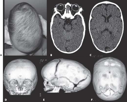
Abdulrahman J. Sabbagh, Jeff rey Atkinson, Jean-Pierre Farmer, and José Luis Montes
Clinical Presentation
- A 3-month-old child presents with an abnormal head shape since birth.
- He was born at term via a normal vaginal delivery.
- The father has a similar but more accentuated head shape.
- The physical examination is otherwise normal.
- A computed tomography (CT) scan of the head is done and shown in Fig. 68.1.

Fig. 68.1 (A) Head photograph, (B,C) axial computed tomography (CT) scan, and (D) three-dimensional reconstructed CT scan, (E,F) of a child with craniosynostosis.
Questions
- Describe the head shape and the CT scan. What is the diagnosis?
- What is scaphocephaly?
- How common is scaphocephaly?
- What is the prognosis of scaphocephaly?
- What is the risk of developing hydrocephalus in patients diagnosed with craniosynostosis? What is the risk for this patient?
- What are some treatment options and their possible related morbidities?
- What is the accepted hypothesis for the pathogenesis of deformities caused by craniosynostosis?
- What is more clinically important, timing of head and brain growth or timing of normal sutural closure, and why?
Answers
- Describe the head shape and the CT scan. What is the diagnosis?
- The photograph, plain CT, and three-dimensional reconstructed CT scans show an elongated boatlike skull with a closed sagittal suture (Fig. 68.1).
- Also seen is frontal and occipital bossing.
- The diagnosis is scaphocephaly.
- What is scaphocephaly?
- Scaphocephaly can be described as a boat-shaped head, caused by synostosis of the sagittal suture, leading to a long and thin skull.1
- There is bifrontal and occipital symmetric compensatory bossing.
- There is usually a midline ridge, and there may be a saddle deformity where the synostosis began.
- It may be evident at birth and progresses to become more pronounced with time.
- It is the most common type of synostosis and is more common in males than females.
- How common is scaphocephaly?
- Epidemiology of scaphocephaly is described below.
- What is the prognosis of scaphocephaly?
- This deformity, like other forms of craniosynostosis, usually progresses with time and will have an important impact on the growing child’s social and psychological status.3
- These children usually would develop normally from a neurologic standpoint.
- There is a slight risk of increased intracranial pressure (ICP).3
- What is the risk of developing hydrocephalus in patients diagnosed with craniosynostosis? What is the risk for this patient?
- Renier et al. in 1982 measured ICP using epidural sensor in 92 patients for 12–24 hours.4 He found the following results as they relate to ICP.
- In one suture synostosis, 62% were normal, 24% were borderline and 14% were high.
- In several sutures synostosis, 19% were normal, 34% were borderline and 47% were high.
- The incidence of hydrocephalus in sagittal craniosynostosis is remarkably rare (~0.3%), except in patients with prematurity or those who have been shunted.5
- On the other hand, in syndromic craniosynostosis (Crouzon syndrome, Pfeiff er syndrome, Apert syndrome, etc.), the incidence of hydrocephalus may range from 20 to 40%.5–7 The former two are more likely to be associated with shunt dependent hydrocephalus than the latter.7
- Renier et al. in 1982 measured ICP using epidural sensor in 92 patients for 12–24 hours.4 He found the following results as they relate to ICP.
- What are some treatment options and their possible related morbidities?
- Ideally, surgical repair should be done at 2–3 months of age. When done before 3 months of age, the following options are available:
- Midline strip craniectomy8
- A 4–8 cm wide craniectomy is performed, from the coronal to just posterior to the lambdoid sutures.
- Bilateral barrel stave osteotomies are then done (Fig. 68.2).
- Blood transfusion is common.
- There is up to a 10% chance of having a residual bone defects.
- Good cosmetic results can be obtained (Fig. 68.3).
- Pi procedure9,10
- This procedure immediately provides anteroposterior shortening of the skull.
- It may also give a better cosmetic result.
- Endoscopic strip with molding helmet therapy
- This method significantly lowers the rate of transfusion.11
- In cases where treatment is contemplated late (after 6 months of age), the following options are available.
- What is the accepted hypothesis for the pathogenesis of deformities caused by craniosynostosis?
- An older hypothesis described by Virchow (in 1851) states, “Craniosynostosis is caused by a lack of growth perpendicular to the fused suture and compensatory growth parallel to the suture in the calvarial vault.”13
- The currently accepted hypothesis devised by Delashaw et al. states, “The calvarial bones directly adjacent to a fused suture act as a single bone plate with decreased growth potential.”13
- Asymmetrical bone deposition occurs at the sutures along the perimeter of the bone plate with increased bone deposition at the outer margin.13
- Nonperimeter sutures in line with the fused suture deposit bone symmetrically at their sutural edges.13
- Perimeter sutures adjacent to the fused suture compensate to a greater degree than other distant sutures.13
- What is more clinically important, timing of head and brain growth or timing of normal sutural closure, and why?
- Timing of head and brain growth is more clinically significant than normal suture closure.1
- The brain doubles in size by 6 months of age and doubles again by 2 years of age.
- The skull is 35% of its adult size at birth, and 90% of the adult size is reached by age 7 years.
- Timing of head and brain growth is more clinically significant than normal suture closure.1
Only gold members can continue reading. Log In or Register to continue
Related
Related posts:
