69 Spontaneous Cerebrospinal Fistula

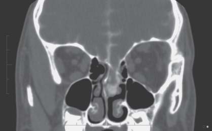
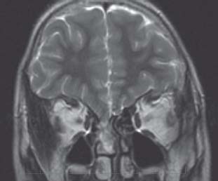
Case 69 Spontaneous Cerebrospinal Fistula


Jeffrey Atkinson, José Luis Montes, and Abdulrahman J. Sabbagh


Image Clinical Presentation


  • A 5-year-old boy presents with his third episode of bacterial meningitis.
  • The child has a history of prematurity with a mild hemiparesis and mild developmental delay.
  • There is no history of trauma.
  • Physical examination reveals no skin defects or cutaneous markers.
  • On detailed questioning, the child does recall that his nose is frequently “runny,” and with prolonged forward positioning small amounts of clear fluid can be found dripping from the nose.

Image Questions


Image

Fig. 69.1 Computed tomography coronal scan through the anterior fossa and ethmoids, with infusion of metrizamide intrathecal contrast.


Image

Fig. 69.2 T2-weighted coronal magnetic resonance image through the anterior cranial fossa.


Image Answers


  1. What are the potential causes of spontaneous CSF fistula?

  2. Where are the potential sites of CSF leak?

    • CSF leak may occur in any area with one of the above pathologies, but obviously occult leaks into the nasal cavity or from the spine into the epidural or lumbar fascial compartments may occur and may be difficult to detect.

  3. What other clinical syndromes might result from CSF fistula?

    • CSF fistula might result in CSF infection as in this case. Any chronic communication between the environment and meningeal space may produce acute, recurrent or chronic meningitis or other central nervous system (CNS) infections.
    • Low-pressure headache may also be attributable to a chronic CSF leak.2
    • Finally, superficial hemosiderosis has been reported following chronic CSF fistula into a traumatic pseudomeningocele with neovascularization and repeated hemorrhage. This could obviously present with mental deterioration, hearing, balance loss, and other cranial nerve deficits.3,4

  4. What would be the diagnostic tests indicated in this patient?

    • In a patient where CSF fistula is suspected, an MRI scan of the complete neuraxis would be imperative. In some instances this may not be diagnostic.2
    • A CT scan can be helpful to demonstrate bone defects. Cisternal infusion of contrast media followed by a thin-cut CT, might be able to demonstrate extra CNS flow of fluid.2
    • In rare instances, a nuclear medicine study with lumbar cisternal infusion of radionucleotide tracer may also be used to demonstrate a small or slow communication, though with less anatomic detail.1

  5. Describe the findings on the scans.

    • The coronal T2-weighted MRI scan and the coronal reconstruction of the postcisternal infusion of contrast CT scan both show a defect in the ethmoidal bone of the frontal cranial fossa with herniation of tissue and CSF through the defect into the nasal cavity.

  6. Describe at least two surgical approaches for repair of the above lesion.

    • There are essentially two surgical approaches to this lesion.

      • A bifrontal craniotomy with repair of the defect from above using bone, muscle graft, and a vascularized pericranial flap would be possible. This might be possible entirely with an extradural approach, but an intradural inlay graft might protect better against CSF leak.1,2
      • Alternatively, there is significant experience in some centers with an entirely endoscopic intranasal approach to the repair of this lesion with again vascularized mucosal flap and an inlay graft if possible.5
< div class='tao-gold-member'>

Only gold members can continue reading. Log In or Register to continue

Stay updated, free articles. Join our Telegram channel

Jul 16, 2016 | Posted by in NEUROSURGERY | Comments Off on 69 Spontaneous Cerebrospinal Fistula

Full access? Get Clinical Tree

Get Clinical Tree app for offline access