74 Hemifacial Spasm and Microvascular Decompression

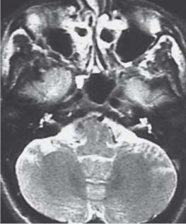
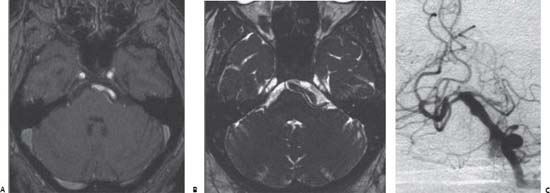
Case 74 Hemifacial Spasm and Microvascular Decompression


Bassem Sheikh


Image Clinical Presentation


  • A 35-year-old man presents with a 10-year history of spasmodic twitching on the left side of his face.
  • The twitching involves the left eyelid and causes intermittent spasmodic closure of the eye.
  • The attacks of hemifacial spasm (HFS) are exacerbated by stress.
  • Clinical examination did not reveal any neurologic deficit.


Image

Fig. 74.1 T2-weighted magnetic resonance image at the level of the posterior fossa.


Image

Fig. 74.2 (A) T1- and (B) T2-weighted magnetic resonance images at the level of the posterior fossa and cerebellopontine angle. (C) Vertebral digital subtraction angiography of the same patient.


Image Questions


  1. What is the most likely diagnosis? The differential diagnosis?
  2. Describe the pathophysiologic basis of HFS.
  3. Interpret the magnetic resonance images (MRIs) shown in Fig. 74.1 (which represent the current patient’s findings) and Fig. 74.2 (which represent another patient’s findings, with the same diagnosis).
  4. What is the underlying cause of HFS?
  5. What are the surgical and nonsurgical therapeutic options?
  6. Briefly describe your surgical procedure for HFS.
  7. What is the expected surgical outcome?
  8. What are the possible surgical complications?

Image Answers


  1. What is the most likely diagnosis? The differential diagnosis?

  2. Describe the pathophysiologic basis of hemifacial spasm.

    • HFS is a neuromuscular disorder that is characterized by paroxysmal bursts of involuntary, intermittent, or continuous clonic movements that progress to sustained tonic activity occurring in the muscles innervated by the facial nerve.
    • HFS represents a segmental myoclonus of muscles innervated by the facial nerve.
    • Irritation of the facial nerve nucleus is believed to lead to hyperexcitability of the facial nerve nucleus, whereas irritation of the proximal nerve segment may cause ephaptic transmission within the facial nerve.
    • Either mechanism explains the rhythmic involuntary myoclonic contractions observed in HFS.
    • The disorder presents usually unilaterally, although bilateral involvement may occur rarely in severe cases.
    • Typically, hemifacial spasm results secondary to vascular cross-compression of the myelinated facial nerve at or proximal to the junctional area of central and peripheral myelin (the root exit zone) of the nerve.1

  3. Interpret the MRIs shown in Fig. 74.1 (which represent the current patient’s findings) and Fig. 74.2 (which represent another patient’s findings with the same diagnosis).

    • Figure 74.1 reveals a T2-weighted MRI of the posterior fossa at the level of the internal auditory meatus. It reveals a flow void structure representing a blood vessel that is crossing the facial root exit level.
    • Figure 74.2 represents another patient with hemifacial spasm. T1 – and T2-weighted MRIs of the posterior fossa at the level of the cerebellopontine angle (CPA). Note the tortuous basilar artery that is compressing the left CPA at the facial exit root.
    • The same patient had a digital subtraction angiography of the vertebral artery (Fig. 74.2C) that confirmed the presence of dolichoectasia of the basilar artery. Note that not all HFS cases are idiopathic. The aim of investigating the patient is to rule out any underlying pathology.

  4. What is the underlying cause of HFS?

    • The actual cause of HFS is debatable.
    • Most cases of HFS are caused by an ectatic blood vessel that irritates the facial nerve by compressing or forming a loop around the nerve at the nerve exit zone. The usual offending artery is the posterior inferior cerebellar artery complex, the anterior inferior cerebellar artery, or the vertebral artery.2
    • A minority of cases of HFS are caused by a venous compression.2
    • Rarely, the condition may be secondary to facial nerve injury, facial nerve compression by a CPA tumor, regeneration of the facial nerve following facial palsy, or it may be a result of a brainstem lesion such as a stroke or a multiple sclerosis plaque.

  5. What are the surgical and nonsurgical therapeutic options?

  6. Briefly describe your surgical procedure for hemifacial spasm.

    • Positioning and opening

      • Lateral decubitus position
      • Retromastoid craniectomy (2.0–2.5 cm in diameter)
      • Dura is incised.
      • Cerebrospinal fluid is drained slowly, allowing the structures of the posterior fossa to fall away without retraction.
      • Lateral or inferolateral cerebellar exposure of the CPA

    • Dissection and decompression

      • The acousticofacial bundle is identified.
      • The facial nerve may be stimulated for verification.
      • The offending vessel is identified, and using micro-dissection and gentle manipulation, the adhesions and compressions from the vessel(s) on the facial nerve are lysed, and the nerve and vessel(s) are freed from one another.
      • Small implants of shredded Teflon felt are placed to hold the vessel away from the cranial nerve root exit zone by changing the axis of the loop. Other techniques may include performing a dural sleeve to hold the artery away from the nerve or gluing the artery to the posterior fossa dura.
      • Veins are treated similarly or coagulated and divided.3,4

  7. What is the expected surgical outcome?

    • Generally, patient should be informed of a possible nonresponse.
    • Excellent results (complete or nearly complete abolition of spasm) are expected in three-quarters of patients at 1 month after operation.
    • Long-term follow-up reveals more patients with total relief of their spasm.3,4
    • Patients having reoperation should expect lower results: 61% complete or nearly complete abolition of spasm.4
    • If the patient is still having spasm in the postoperative period, conservative follow-up will usually show progressive resolution of the residual spasm within the following month.

  8. What are the possible surgical complications?

    • General complication related to posterior fossa surgery
    • Specific to facial nerve microvascular decompression:

      • Partial or complete, temporary or permanent facial palsy may result from manipulation of the facial nerve.
      • Owing to the immediate proximity of the eighth cranial nerve, microvascular decompression of the facial nerve for hemifacial spasm has a risk of producing ipsilateral hearing loss of various degrees. This may result from stretching the eighth cranial nerve between its exit from the brainstem and its entry into the internal auditory meatus as the surgeon places cerebellar retraction to expose the facial nerve root exit. This complication may be controlled if intraoperative brainstem auditory evoked potentials are monitored.3,4
< div class='tao-gold-member'>

Only gold members can continue reading. Log In or Register to continue

Stay updated, free articles. Join our Telegram channel

Jul 16, 2016 | Posted by in NEUROSURGERY | Comments Off on 74 Hemifacial Spasm and Microvascular Decompression

Full access? Get Clinical Tree

Get Clinical Tree app for offline access