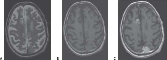
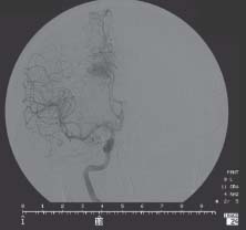
78 Stereotactic Radiosurgery Case

Case 78 Stereotactic Radiosurgery Case


Carmina M. Angeles and Dennis G. Vollmer


Image Clinical Presentation


Image Questions


  1. Interpret the MRI (Fig. 78.1).
  2. What are the differential diagnoses?
  3. What is the next step in management?
  4. You decide to obtain a four-vessel angiogram (Fig. 78.2). Interpret the images.
  5. What are the management options for the lesion seen on angiography?
  6. What are the management options for this patient?

    This patient elected to undergo stereotactic radiosurgery (SRS) to both lesions. The treatment was planned for a dose of 22 Gy at 50% isodose line to the frontal arteriovenous malformation (AVM), and 16 Gy to the 50% isodose line for the dural based parietooccipital lesion.


  7. Describe the main steps involved in the planning and completion of SRS.
  8. What are the outcomes of SRS on AVMs?
  9. What are the outcomes of SRS on meningiomas?
  10. What are the complications of SRS?


Image

Fig. 78.1 Axial magnetic resonance images of the brain: (A) T2-weighted, (B) T1-weighted, and (C) T1-weighted with contrast.


Image

Fig. 78.2 Cerebral angiogram, left internal carotid injection, anteroposterior view.


Image Answers


  1. Interpret the MRI (Fig. 78.1).

    • Figure 78.1A and B reveal noncontrasted T1- and T2-weighted images depicting two lesions. There is an ~2.6-cm nonhemorrhagic right frontal parafalcine lesion with several large flow voids.
    • There is a second nonhemorrhagic hypodense lesion on the T1-weighted image ~1.4 × 3.3 × 2.4-cm located in the left parietooccipital area.
    • In the postgadolinium T1-weighted image in Fig. 78.1C, the right frontal lesion is heterogeneously enhancing, whereas the left parietooccipital lesion is more homogeneous and appears to be dural-based.

  2. What are the differential diagnoses?

    • Differential diagnoses for the right frontal lesion include

      • Arteriovenous malformation (AVM)
      • Cavernous malformation
      • Hemangiopericytoma

    • Differential diagnoses for the left parietooccipital lesion include

      • Meningioma (most likely)
      • Metastases
      • Other less likely lesions: hemangiopericytoma, lymphoma, brain abscess1

  3. What is the next step in management?

    • A cerebrovascular study such as four-vessel cerebral angiography, magnetic resonance angiography, or computed tomography angiography is warranted to assess the frontal lesion. The patient did undergo in this case a cerebral angiogram.
    • In addition, an electroencephalogram (EEG) should be performed to help localize the source of the seizure. The results of the EEG, however, in this case were essentially of a normal awake person.
    • A lumbar puncture to rule out cerebrospinal fluid xanthochromia may also help delineate whether the seizure was a result of a hemorrhage from the likely AVM.

  4. You decide to obtain a four-vessel angiogram (Fig. 78.2). Interpret the image.

    • Figure 78.2 reveals an angiogram, right carotid artery injection, showing an AVM feeding primarily from the right pericallosal artery and appears to be draining into a right frontal cortical vein.
    • Considering the size of the lesion being less than 3 cm, location in noneloquent brain, and a superficial draining vein, this lesion is a grade I based on the Spetzler–Martin grading system (please refer to Case 28 for AVM grading).2

  5. What are the management options for the lesion seen on angiography?

    • There are essentially three treatment options for an AVM: surgery, embolization, and SRS. In addition, preoperative embolization followed by surgical resection may be another option that significantly reduces the risks of surgery and can be curative compared with embolization alone.24

  6. What are the management options for this patient?

    • Treatment options for this patient with an AVM include various combinations of open surgical resection, embolization, SRS, and observation alone, as described above.
    • Treatment options for the dural-based lesion in the parietooccipital lobe include surgical resection, biopsy, radiosurgery, or observation alone.
    • A combination of treatment modalities is possible in this case, based on the patient’s preference, age, comorbidities, and other medical conditions.
    • Given the advanced age of the patient, the small size of the lesions and the convincing appearance of the lesions on MRI and angiography, some may argue that SRS alone (without having tissue biopsy specimen for pathologic diagnosis) may be a valid option in this particular case.5

  7. Describe the main steps involved in the planning and completion of SRS.

    • SRS is a minimally invasive treatment technique used to deliver a focal dose of high-energy radiation to a target. This target is localized stereotactically using three-dimensional image processing. Normal surrounding tissues and structures are spared.6
    • A Leksell stereotactic coordinate frame is placed under local anesthetic agent and is centered on the target (tumor or AVM).6 Other localizing frames of reference may also be used.
    • MRI or CT scan of the brain is performed and the images are then used for treatment planning with an interactive computer, which provides information on the volume of the target, dose of radiation, dose to volume ratio, isodose curves, etc.5,6
    • There are two predominant types of delivery devices for SRS6:

      • Gamma knife (GKS)

        • A radioactive isotope such as Cobalt 60 is focused on the target by interchangeable helmets with different size collimators via 201 sources.
        • The additive effect of multiple isocenters of radiation results in a high dose of radiation delivered in a conformal manner.5

      • Linear accelerator (LINAC)

        • By moving a single beam of radiation in arcs around a patient’s head, the amount of radiation and the size of the beam can also be collimated and tailored to the target of interest.5

    • Steroids may be concomitantly given if there is a concern about brain edema or eloquent neural tissue damage.

  8. What are the outcomes of SRS on AVMs?

    • 5-year AVM obliteration rate ranges from 66 to 89%.7, 8

      • In GKS cases, it is ~72 to 89%
      • In LINAC cases, it is ~60%

  9. What are the outcomes of SRS on meningiomas?

    • 5-year tumor control rate was 90 to 98%.5,911
    • In a series of 400 cases, tumor volume decreased in ~70%, remained the same in ~28%, and increased only in ~2.5%.10
    • Symptomatic complications were limited to ~5%.9

  10. What are the complications of SRS?

    • Perilesional edema in 7 to 16%10,12
    • Damage to critical eloquent brain structures such as the optic nerves or other cranial nerves12
    • After SRS hemorrhage during the time before obliteration of the AVM, the rate is between 6 and 13%.7
    • Other potential complications include headaches, seizures, intracranial cysts, 13 and radiation necrosis.14
< div class='tao-gold-member'>

Only gold members can continue reading. Log In or Register to continue

Stay updated, free articles. Join our Telegram channel

Jul 16, 2016 | Posted by in NEUROSURGERY | Comments Off on 78 Stereotactic Radiosurgery Case

Full access? Get Clinical Tree

Get Clinical Tree app for offline access