85 Compression Fracture of the Thoracolumbar Spine

Case 85 Compression Fracture of the Thoracolumbar Spine


Eric P. Roger and Edward Benzel


Image

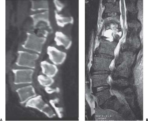
Fig. 85.1 (A) Sagittal reconstructed computed tomography of the lumbar spine and (B) sagittal T2-weighed magnetic resonance image of the lumbar spine are shown.


Image Clinical Presentation


  • A 49-year-old man presents with back pain.
  • He has a previous history of a fall ~3 months ago in the shower. He had no back pain prior to the fall.
  • His past medical history is remarkable for arthroscopic knee surgery a few weeks prior to the fall, complicated by postoperative infection leading to sepsis. He was on intravenous (i.v.) antibiotics for 3 months.
  • He denies any bladder or bowel dysfunction.
  • On physical examination, he is neurologically intact and nonmyelopathic.

Image Questions


  1. Please interpret the images in Fig. 85.1.
  2. What do you think the cause of this fracture might be?
  3. Assuming the fracture may be pathologic, what would be the differential diagnosis for the underlying pathology?

    You are concerned about possible osteomyelitis. You obtain a computed tomography (CT) guided aspiration/biopsy of this lesion. Gram stain, final cultures, and pathology are all negative.


  4. Can you rule out osteomyelitis as an underlying cause for this fracture?
  5. How would osteomyelitis affect your surgical decision making?
  6. What is the retropulsed bone compressing? Why does it matter?
  7. What are the therapeutic options for this patient? Describe the available surgical approaches.

    The patient tried a thoracolumbar spinal orthosis brace. However, when standing he experienced excruciating pain. Standing radiographs revealed progression of the kyphosis from 30 degrees to more than 60 degrees.


  8. What are your surgical objectives?
  9. What stabilization constructs are available to you?
  10. How do you classify thoracolumbar fractures? How do you classify this fracture?
  11. What is the mechanism for this fracture?


Image

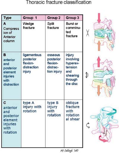
Fig. 85.2 Classification of thoracic fractures : (A) Type A, injury caused by compression of the anterior column; (B) type B, injury of the anterior column and the two posterior columns with distraction of the anterior or posterior elements; (C) type C, rotational injury of all three columns.

ImageAnswers


  1. Please interpret the images in Fig. 85.1.

    • The most prominent finding is a destruction and/or a burst fracture of the L1 vertebra with involvement of the lower half of the T12 vertebral body.
    • This results in an anterior collapse, with kyphotic angulation deformity in the range of 30 degrees in these supine images.
    • There is evidence of retropulsed bony fragments into the spinal canal.
    • The T2-weighted magnetic resonance image demonstrates hyperintense signal within the T12 and L1 vertebral body, extending into the L1–L2 disk.

  2. What do you think the cause of this fracture might be?

    • Although the patient reports that the pain initiated from a fall in the shower and therefore sounds traumatic, there might be an underlying pathology predisposing this bone to fracture.
    • The patient reported concordant sepsis from an infected knee. Underlying osteomyelitis should be suspected.

  3. Assuming the fracture may be pathologic, what would be the differential diagnosis for the underlying pathology?

    • Differential diagnosis includes1

      • Infectious

        • Bacterial seeding from sepsis
        • Tuberculosis

      • Neoplastic

        • Primary bone tumor (myeloma common)
        • Metastases, lymphoma

      • Osteoporosis
      • Traumatic

  4. Can you rule out osteomyelitis as an underlying cause for this fracture?

    • No, this cannot be entirely ruled out. This patient has been on i.v. antibiotics for 3 months. Although the biopsy or aspiration is negative, this may certainly have been an osteomyelitis initially that has now been “sterilized” by the antibiotics.
    • It has been reported that radiologic findings often “lag behind” clinical improvement and blood markers (such as C-reactive protein, erythrocyte sedimentation rate, and white blood cell count).
    • Furthermore, the lack of growth on culture does not necessarily prove that this collection is sterile. The long-term use of antibiotics may preclude culture growth, even though bacteria may still be present.24

  5. How would osteomyelitis affect your surgical decision making?

    • Acute vertebral osteomyelitis is rarely initially treated surgically unless there is neural compromise, significant progressive deformity, or intractable pain. An aggressive course of antibiotic therapy is usually indicated (for 2 to 3 months). 2
    • As previously mentioned, the images often “lag behind.” Furthermore, successfully treated vertebral osteomyelitis will often progress to spontaneous fusion.
    • The issue with treating vertebral osteomyelitis surgically in the acute setting is one of spinal hardware contamination, although this can certainly be managed if surgery is clinically necessary.57
    • Once the osteomyelitis has been successfully “sterilized,” as suspected in this patient, surgical stabilization may then be required for ongoing pain, instability, or new neurologic compromise.
    • One may consider continuing the i.v. antibiotics postoperatively for 6 weeks or more.

  6. What is the retropulsed bone compressing? Why does it matter?

    • On Fig. 85.1B, it appears that the retropulsed bone is compressing the thecal sac below the level of the conus medullaris, thus affecting the cauda equina. The conus is most commonly located at L1, but may often terminate at T12.
    • The patient’s lack of myelopathic findings suggests that the conus is above the retropulsed bony fragment in this case.
    • Whether the retropulsed bone is compressing the conus has a large impact on the proposed risks of the surgery, choice of approach, and ease of thecal sac decompression intraoperatively.

  7. What are the therapeutic options for this patient?
    Describe the available surgical approaches.


    • Therapeutic options include

      • Conservative management: bracing2
      • Surgical decompression and stabilization37

        • Iliac crest autograft should be harvested prior to exposing the infected level. Measurements are taken preoperatively from the CT scan.
        • Anterior approach alone8: T12 and L1 corpectomy with intervertebral cage placement ± anterolateral plating. This would involve combined left-sided transthoracic/flank retroperitoneal approach with diaphragmatic take-down. (Note: This is a difficult approach and the assistance of an “exposure” surgeon—general or vascular—is recommended.)
        • Anterior and posterior approach (combined): supplementation of the above approach with posterior pedicle fixation, one to three levels above and below
        • Posterior approach alone9,10: transpedicular, costotransversectomy, or lateral extracavitary approach to corpectomy and cage, supplemented by pedicle screw fixation. (Note: The T12-L1 area is a junctional level; as such, it is recommended to proceed with at least two-level fixation above and below the level of involvement.)

      • Kyphoplasty is not a valid option in this case.

  8. What are your surgical objectives?

    • Surgical objectives4

      • Neural decompression from the retropulsed bony fragment
      • Restoration of alignment (i.e., correction of the kyphosis)
      • Restoration of anterior weight-bearing capacity (anterior and middle columns)
      • Fixation and fusion (arthrodesis and instrumentation)

  9. What stabilization constructs are available to you?

    • Anterior constructs include4,8

      • Structural autograft or allograft such as iliac crest or tibial strut
      • Artificial cage (titanium or polyetheretherketone) filled with autograft, allograft or ceramics (β-tricalcium-phosphate [β-TCP])
      • Expandable cages may offer an advantage in reducing the kyphosis by distracting the anterior column.
      • Anterolateral plate and screw construct

    • Posterior constructs include4

      • Pedicle screw fixation
      • Laminar hooks
      • Percutaneous pedicle screw fixation may be attractive if posterior decompression is not indicated (i.e., pedicle screws as a “backup”).
      • Bone grafting with autograft, allograft, or β-TCP

  10. How do you classify thoracolumbar fractures? How do you classify this fracture?

    • Fracture classification (simplified)11,12 (Fig. 85.2)

      • Type A: Vertebral body compression

        • A1—Impaction fractures (wedge)
        • A2—Split fractures
        • A3—Burst fractures

      • Type B: Anterior and posterior element injury with distraction

        • B1—Posterior disruption predominantly ligamentous (ligamentous Chance injury)
        • B2—Posterior disruption predominantly osseous (bony Chance injury)
        • B3—Anterior disruption through disk (hyperex-tension shear injury)

      • Type C: Anterior and posterior element injury with rotation

        • C1—Type A (compression) and rotation
        • C2—Type B and rotation
        • C3—Rotational shear injuries

    • This fracture would classify as an A3 (anterior column only, with burst features).

  11. What is the mechanism for this fracture?

    • Mechanism: Axial compression with angular flexion moment arm.
    • There is no posterior element distraction associated to it.
    • There likely is underlying bone fragility from previous osteomyelitis, although this cannot be confirmed.
< div class='tao-gold-member'>

Only gold members can continue reading. Log In or Register to continue

Stay updated, free articles. Join our Telegram channel

Jul 16, 2016 | Posted by in NEUROSURGERY | Comments Off on 85 Compression Fracture of the Thoracolumbar Spine

Full access? Get Clinical Tree

Get Clinical Tree app for offline access