88 Anterior versus Posterior Approach to the Cervical Spine

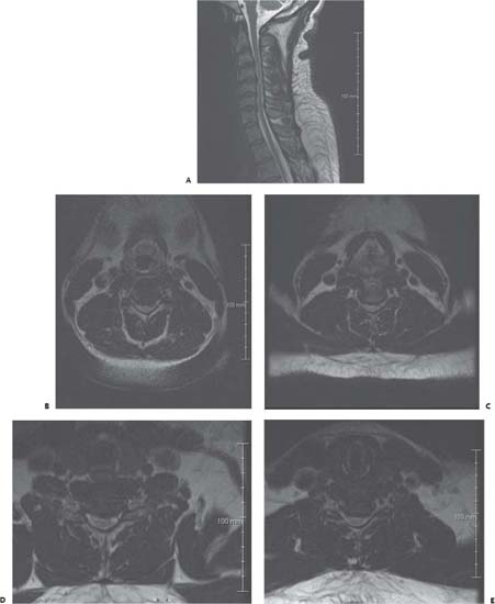
Case 88 Anterior versus Posterior Approach to the Cervical Spine


Amgad S. Hanna and Remi Nader


Image

Fig. 88.1 Cervical spine T2-weighted magnetic resonance images with (A) midsagittal section and axial sections through (B) C3–C4, (C) C4–C5, (D) C5–C6, and (E) C6–C7, respectively.


Image Clinical Presentation


Image Questions


  1. Interpret the MRI scan.
  2. What is your management?
  3. What factors will favor an anterior versus a posterior surgical approach?
  4. Postoperatively, the patient does not improve (no obvious intraoperative complications). What do you tell the patient?

ImageAnswers


  1. Interpret the MRI scan.

    • Cervical canal stenosis is seen extending from C3 to C7 with multiple-level disk bulges, more so at C4–C5.
    • The sagittal images do not fully account for the narrowing seen in the axial cuts.
    • There is increased signal intensity of the cord on T2-weighted MRIs demonstrating damage to the spinal cord, likely from the stenosis.
    • The region of the posterior longitudinal ligament (PLL) is thickened and hypointense. This could be ossified PLL (OPLL); obtaining a computed tomography (CT) scan would confirm this finding, if present.
    • No bony changes or extradural tissues are seen to suggest the diagnosis of epidural abscess, which could well be a possibility in an HIV scenario. HIV can also lead to neuropathic changes and changes within the cord.

  2. What is your management?

    • This patient is myelopathic in the context of significant cervical stenosis; therefore, surgery is the most appropriate treatment measure in this case.
    • In the meantime, an infectious workup for HIV should be performed (including CD4 count, viral load, etc.). Acquired immunodeficiency syndrome-(AIDS)-related myelopathy should also be in the differential diagnosis.

  3. What factors will favor an anterior versus a posterior surgical approach?

  4. Postoperatively, the patient does not improve (no obvious intraoperative complications). What do you tell the patient?

    • The main goal of surgery is to stop further deterioration.
    • Improvement rate of neurologic function is ~50% (ranging from 37 to 85%) in myelopathic patients; therefore, it is not unusual to see no change postoperatively.68
    • Also, he has myelopathic changes within the spinal cord on the MRI, and some of his symptoms may be related to HIV myelopathy and peripheral neuropathy.
    • One management approach, at this stage, would be to continue observation while initiating rehabilitative measures such as physical therapy.
< div class='tao-gold-member'>

Only gold members can continue reading. Log In or Register to continue

Stay updated, free articles. Join our Telegram channel

Jul 16, 2016 | Posted by in NEUROSURGERY | Comments Off on 88 Anterior versus Posterior Approach to the Cervical Spine

Full access? Get Clinical Tree

Get Clinical Tree app for offline access