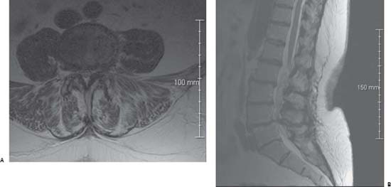
91 Cauda Equina Syndrome

Case 91 Cauda Equina Syndrome


Cristian Gragnaniello and Remi Nader


Image

Fig. 91.1 (A) Axial T2- weighted magnetic resonance image (MRI) at the L5 pedicle level. (B) Sagittal T2-weighted MRI through the lumbar spine.


Image Clinical Presentation


Image Questions


  1. What is the clinical diagnosis? Describe the main clinical features of this diagnosis.
  2. What conditions can cause these symptoms and signs?
  3. Interpret the MRI findings.
  4. What, if any, other imaging studies would you obtain?
  5. How do you manage this condition?
  6. What is the likelihood of her recovering bladder function?
  7. Describe bladder micturition physiologic mechanisms.
  8. If there is no relief from the pain, what is your workup and management?

Image Answers


  1. What is the clinical diagnosis? Describe the main clinical features of this diagnosis.

    • Chronic cauda equina syndrome1
    • The fully developed syndrome consists of many clinical symptoms and signs that can be present in various degrees in each individual patient. This can sometimes lead to a delay in the diagnosis that can influence the outcome (Table 91.1).24

  2. What conditions can cause these symptoms and signs?

    • Causes of onset of this complex syndrome must always be kept in mind when approaching the patient. They are summarized in Table 91.2.5

  3. Interpret the MRI findings.

    • Grade 1 degenerative spondylolisthesis at L4/5 causing severe narrowing of the canal and nerve root impingement.6
    • The nerve root impingement correlates very well with the clinical signs of radiculopathy.7,8
    • A sign that the spondylolisthesis is due to degenerative changes can be found in the sagittal image that shows a malalignment of the spinous process of L4 that displaces with the vertebral body due to maintenance of the integrity of the neural arch.6
    • The filamentous-looking structures above L4 are just the nerve roots; they may appear that way in cases of severe stenosis.
    • Marked thickening of the ligamentum flavum is seen in the axial image.

  4. What, if any, other imaging studies would you obtain?

    • To assess the motion of the lumbar segments that are studied, dynamic radiographs done with flexion and extension should be obtained to rule out instability.9,10
    • A computed tomography scan to can determine whether there is a pars defect and to assess the orientation and size of the pedicles, to study the central canal, the lateral recess, and the foramen.11
    • Radiographs of the thoracic and lumbar spine and MRI of the whole spine may be also obtained because even if the symptoms suggest the cauda equina syndrome, there may be a more proximal lesion (this is especially important in cases where the stenosis is mild and does not fully account for the symptoms).12

  5. How do you manage this condition?

    • Decompressive bilateral laminectomies using a high-speed drill and acorn or diamond burr to avoid any possible other manipulation of the roots
    • The extent of decompression should allow access laterally to the thecal sac at the level of L4–L5.1
    • Timing of the surgery is essential (semiurgent, within 48 hours).1,3,5,1216
    • Fusion must follow if there are instability signs on imaging. Some will elect to fuse anyway, given the significant bilateral decompression necessary here and the likely resection of the disk.17

  6. What is the likelihood of her recovering bladder function?

    • Less than 50% in view of one-month history of dysfunction.
    • The recovery will be better compared with patients who suffered from a complete bladder paralysis14
    • Early surgical intervention within 48 hours is essential. In general, the outcome is much better compared with those operated on after 48 hours.4,1416,18

  7. Describe bladder micturition physiologic mechanisms.

    • There are two phases: bladder filling and emptying (Fig. 91.2).
    • Bladder-filling phase19

      • The bladder accumulates increasing volumes of urine.
      • The pressure within the bladder must be lower than the urethral pressure during the filling phase.
      • Bladder filling is dependent on the intrinsic visco-elastic properties of the bladder and inhibition of the parasympathetic nerves.
      • Sympathetic nerves facilitate urine storage.
      • Sympathetic input to the lower urinary tract is constantly active during bladder filling.
      • Pudendal nerve becomes excited.

    • Bladder-emptying phase19

      • A voluntary signal is sent from the brain to begin urination and continues until the bladder is empty.
      • Bladder afferent signals ascend the spinal cord to the periaqueductal gray.
      • These afferents project to the pontine micturition center and to the cerebrum.20
      • Once the voluntary signal to begin voiding has been issued, neurons in pontine micturition center fire maximally.
      • This causes excitation of sacral preganglionic neurons leading to the wall of the bladder to contract.
      • The pontine micturition center also causes inhibition of Onuf’s nucleus, leading to relaxation of the external urinary sphincter.21

    • In cases of incontinence related to cauda equina

      • In cauda equina syndrome, afferent and efferent nerves are both lesioned, and the bladder can become flaccid and distended temporarily.
      • The detrusor muscle gradually becomes spontaneously active, with intermittent contractions that may cause dribbling.
      • The bladder then shrinks and its wall hypertrophies.
      • This mechanism is called denervation hypersensitization.

  8. If there is no relief from the pain, what are your workup and management?

    • In this situation, to treat refractory residual pain after all conservative therapies failed, functional neurosurgical techniques can yield good results in many cases.
    • Different procedures are available and can be considered, including

      • Spinal cord stimulation
      • Ablative procedures like dorsal root entry zone (DREZ) lesioning (or DREZotomy)22,23
< div class='tao-gold-member'>

Only gold members can continue reading. Log In or Register to continue

Stay updated, free articles. Join our Telegram channel

Jul 16, 2016 | Posted by in NEUROSURGERY | Comments Off on 91 Cauda Equina Syndrome

Full access? Get Clinical Tree

Get Clinical Tree app for offline access