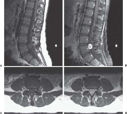
93 Intradural Spinal Tumor

Case 93 Intradural Spinal Tumor


Adam Sauh Gee Wu and Stephen J. Hentschel


Image

Fig. 93.1 Magnetic resonance images of the lumbar spine. (A) Sagittal T1-weighted image, (B) sagittal T1-weighted image with gadolinium, (C) axial T1-weighted image, (D) axial T1-weighted image with gadolinium through the lesion at L5.


Image Clinical Presentation


  • A 25-year-old woman presented with a 1-year history of persistent bilateral leg pain radiating from the low back to the posterior thigh and lateral ankle.
  • The pain is more severe on the left side than it is on the right.
  • Physical examination reveals decreased pinprick sensation over the left lateral leg and ankle. Otherwise, she is neurologically intact.
  • A noncontrast computed tomography (CT) scan of the lumbar spine was obtained and was within normal limits.

Image Questions


  1. Is further imaging necessary? What are you looking for?
  2. A magnetic resonance imaging scan is obtained (Fig. 93.1). Describe the findings and exact location of the abnormality.
  3. What is your differential diagnosis?
  4. What are the indications for and goals of surgery in this case? Is there any role for nonsurgical management?
  5. Describe the surgical procedure you would perform. What intraoperative findings would you expect with respect to the relationships to neural structures and the site of origin of the mass?
  6. What operative adjuncts might you consider?
  7. What are the potential complications of surgery in this case, and what can you do during surgery to reduce the risk of complications?
  8. Hematoxylin and eosin stains of the specimen at low and high power are shown (Fig. 93.2). Describe the histopathology. What is the diagnosis?
  9. Describe the cytogenetics associated with this tumor.
  10. What is the expected outcome of surgery in this case, and what is the potential for recurrence?


Image

Fig. 93.2 Hematoxylin and eosin stains of pathology specimen: representative (A) low and (B) high power views are shown.


Image Answers


  1. Is further imaging necessary? What are you looking for?

    • Although the CT scan is normal, an MRI scan is needed to rule out intradural pathology.

  2. An MRI scan is obtained (Fig. 93.1). Describe the findings and exact location of the abnormality.

    • There is an intradural mass at the L5 level.
    • It is isodense on T1-weighted images, with intense gadolinium enhancement but no obvious dural tail.

  3. What is your differential diagnosis?

    • The most common intradural neoplasms of the spinal cord and cauda equina with the MRI characteristics seen in this case are meningiomas, schwannomas, neurofibromas, and myxopapillary ependymomas.
    • Metastases, hemangiopericytomas, and lipomas are less likely.13

  4. What are the indications for and goals of surgery in this case? Is there any role for nonsurgical management?

    • The indications for surgery are cytoreduction to relieve mass effect, and to obtain tissue for pathologic analysis.1,4
    • The goal of surgery is complete resection of the tumor with preservation of neurologic function,1 but subtotal resection is preferred over aggressive tumor removal, which risks neurologic injury, as most tumors are benign.
    • Nonsurgical management may be considered if the patient has high surgical risk or limited life expectancy.

  5. Describe the surgical procedure you would perform. What intraoperative findings would you expect with respect to the relationships to neural structures and the site of origin of the mass?

    • The mass can be accessed with a standard L5 laminectomy.
    • Bone may be removed from the pedicles and facets to facilitate exposure in some cases, but that is unlikely to be necessary here.
    • The dura should be opened with preservation of the underlying arachnoid to prevent rapid cerebrospinal fluid (CSF) release, which may obscure the operative field, increase the amount of epidural bleeding, and increase the risk of neurologic injury.
    • The dural edges are tacked back and the arachnoid is opened sharply and clipped to the dura with vascular clips.
    • The expected intraoperative findings will vary with the tumor type.
    • Meningiomas commonly arise lateral or posterior to the neural elements, which are usually pushed aside.2

      • Invasion of neural structures is very rare.
      • If there is no risk of neurologic injury from doing so, the tumor should be removed en bloc to prevent spillage of neoplastic cells.
      • The dural attachment is variably extensive and should be coagulated thoroughly or excised if possible.

    • Schwannomas tend to arise from dorsal nerve roots and come from a single fascicle that can be seen entering and exiting the tumor mass.

      • Surrounding fascicles associated with the tumor’s pseudocapsule can often be dissected free.
      • If not, the tumor may be debulked, leaving the pseudocapsule and adherent fascicles behind.

    • In contrast, neurofibromas often arise from ventral nerve roots and may involve and invade multiple adjacent fascicles.1,2

      • The involved nerve root is usually nonfunctional and frequently must be sacrificed to achieve a gross total resection.

    • Myxopapillary ependymomas generally arise from the conus area5 and are surrounded by lumbosacral nerve roots, requiring piecemeal removal.

  6. What operative adjuncts might you consider?

    • Intraoperative ultrasound can be used to determine the limits of the tumor prior to dural opening.
    • An operating microscope should always be used.
    • Direct electrical stimulation of nerve roots with bipolar stimulation and electromyography may be used to identify important functional roots.
    • Electrophysiological monitoring with somatosensory evoked potentials (sensitivity 87%, specificity 90%) and motor evoked potentials (MEPs) (sensitivity 100%, specificity 75%) can be used to detect potentially reversible neurologic injuries.1
    • A cavitating ultrasonic aspirator can be used to help debulk the interior of the tumor.

  7. What are the potential complications of surgery in this case, and what can you do during surgery to reduce the risk of complications?

    • The risk of neurologic morbidity is 15% and that of mortality is 3%.
    • The risk of neurologic worsening is minimized by avoiding manipulation of the nerve roots, spinal cord, and other neural elements and by utilizing aggressive bony removal when necessary.
    • The CSF spaces should be cleared of blood by thorough irrigation prior to dural closure to reduce the risk of postoperative headache, arachnoiditis, and aseptic meningitis.
    • A watertight dural closure using dural patch grafts when necessary will decrease the risk of CSF leak and pseudomeningocele formation.
    • The risk of spinal instability in cases requiring extensive bone removal can be reduced by concurrent instrumentation and fusion.1

  8. Hematoxylin and eosin stains of the specimen at low and high power are shown (Fig. 93.2) . Describe the histopathology. What is the diagnosis?

    • Multiple spindle-shaped cells with tapering nuclei are seen arranged in compactly associated parallel streams. This is typical of the Antoni A pattern.
    • More loosely textured Antoni B areas are also visible, primarily in the upper left corner of the image.
    • Verocay bodies are apparent in the higher magnification image.
    • These features are all consistent with the diagnosis of schwannoma.5

  9. Describe the cytogenetics associated with this tumor.

    • Alterations of chromosome 22q, including loss of the entire chromosome, LOH of 22q, or loss of function mutations of the NF2 gene found at 22q, are the only consistent genetic alterations found in schwannomas.6

  10. What is the expected outcome of surgery in this case, and what is the potential for recurrence?

    • Significant relief of pain and improvement of neurologic symptoms are seen in 80 to 90% of cases.1,7
    • Schwannomas have a 6 to 12% recurrence rate with a mean time to recurrence of 5 years.
    • Subtotal resections have a recurrence rate of less than 15%.1,2
< div class='tao-gold-member'>

Only gold members can continue reading. Log In or Register to continue

Stay updated, free articles. Join our Telegram channel

Jul 16, 2016 | Posted by in NEUROSURGERY | Comments Off on 93 Intradural Spinal Tumor

Full access? Get Clinical Tree

Get Clinical Tree app for offline access