97 Spinal Arteriovenous Fistula

Case 97 Spinal Arteriovenous Fistula


Pascal M. Jabbour and Erol Veznedaroglu


Image Clinical Presentation


Image

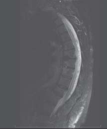
Fig. 97.1 T2-weighted magnetic resonance image (MRI), mid-sagittal section.


Image

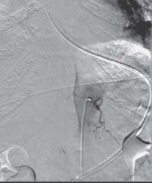
Fig. 97.2 Thoracic spine angiogram, anteroposterior view with selective injection of thoracic artery.


Image Questions


  1. Where do you localize the lesion according to her examination?
  2. Describe the MRI and angiogram findings.
  3. What is the next imaging test to perform?
  4. What are some clinical differences in spinal dural arteriovenous fistulas (DAVFs) and spinal intradural arteriovenous malformations (AVMs)?
  5. What are the different treatment options for this patient?
  6. What are some complications of management of spinal DAVFs?
  7. What are the outcomes of treatment of spinal DAVFs?

ImageAnswers


  1. Where do you localize the lesion according to her examination?

    • She has upper motor neuron signs with increased reflexes, Babinski, and spasticity. Her upper extremities are normal; therefore, the lesion is probably located at the level of her thoracic spine.

  2. Describe the MRI and angiogram findings.

    • The MRI shows edema in the spinal cord, with prominent flow voids representing engorged vessels surrounding the spinal cord.
    • The angiogram demonstrates a DAVF, which is also a type I spinal AVM.

  3. What is the next imaging test to perform?

  4. What are some clinical differences in spinal DAVFs and spinal intradural arteriovenous malformations?

  5. What are the different treatment options for this patient?

    • The goals of treatment consist of interrupting the vein draining the AVF as it penetrates the inner dural layer.2
    • Her options include

      • Open surgery with clipping of the fistula, which usually resides in the sleeve of a nerve root. Laminectomies one level above and one below are usually performed. The dura can be opened in the midline and retraced laterally. The site of the fistula is identified and correlated with imaging studies. The arterialized vein is then coagulated and a limited resection is performed.2,3
      • Endovascular embolization of the fistula4,5

  6. What are some complications of management of spinal DAVFs?

    • Acute neurologic deficit postsurgery or postembolization

      • This necessitates immediate imaging studies via MRI, administration of steroids, followed by possible reexploration.

    • Hematoma
    • Postoperative infection
    • Residual malformation or fistula on postoperative angiography (may require reoperation)

      • It is recommended to perform an angiogram ~1 week after surgical treatment.2

  7. What are the outcomes of treatment of spinal DAVFs?

    • In general, safe and successful treatment is achievable.
    • In most cases, there is improvement of neurologic function or treatment – it usually arrests the progression of the symptoms. These good results can be seen in ~80–90% of patients.1,3
    • Most patients can ambulate after treatment.
    • Bladder symptoms are often improved.
    • Outcome correlates very much with initial neurologic condition upon presentation: patients with less neurologic deficit on presentation will fare better.1,3
< div class='tao-gold-member'>

Only gold members can continue reading. Log In or Register to continue

Stay updated, free articles. Join our Telegram channel

Jul 16, 2016 | Posted by in NEUROSURGERY | Comments Off on 97 Spinal Arteriovenous Fistula

Full access? Get Clinical Tree

Get Clinical Tree app for offline access