Figure 64-1 Cardiovascular autonomic testing in the patient with AAG (left panels) and a normal control (right panels). Heart rate is depicted in green, blood pressure in brown, and respiratory excursions in dark blue. The top panels illustrate a blunted heart rate response to deep breathing. The middle panels illustrate a decreased late phase II, absent phase IV overshoot, and a mildly blunted heart rate response to the Valsalva maneuver (strain phase in light blue). The bottom panels show a mild orthostatic blood pressure drop and marked orthostatic tachycardia during 70-degree head-up tilt (tilt phase in light blue).
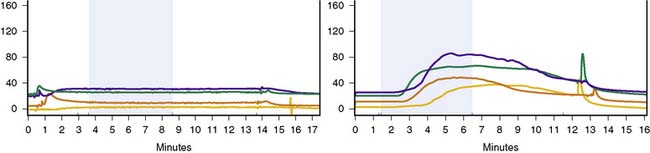
Figure 64-2 Quantitative sudomotor axon reflex test at four sites (forearm-red, proximal leg-blue, distal leg-green, and foot-yellow) in the patient with AAG (left panel) and a normal control (right panel). The time of iontophoresis of acetylcholine (2 mA at each site) is highlighted in light blue. Note the absence of a response in the patient.
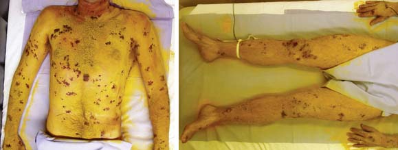
Figure 64-3 Thermoregulatory sweat test (digital photographs). Note the marked anhidrosis except for characteristic small islands of preserved sweating.
CONCLUSION
Our case describes a classic presentation of AAG. AAG represents a pathophysiologically well-described autonomic condition with a heterogenous clinical phenotype beyond such classic presentations. Testing for ganglionic AChR antibodies can help detect unusual presentations of this disorder, which may also be amenable to immunomodulatory therapy even decades after onset of symptoms.
Related posts:
 Malignant Peripheral Nerve Sheath Tumor
Disseminated Sporotrichosis with Multiple Granulomatous Mononeuropathies
Late Sporadic CMT4C—A New KIAA1985 Mutation
Late-Onset Transthyretin Val30Met Familial Amyloid Polyneuropathy Unrelated to Endemic Foci
A Weak, and Numb Patient with Tremor—Antimyelin-Associated Glycoprotein Polyneuropathy
Copper Deficiency Myeloneuropathy
Malignant Peripheral Nerve Sheath Tumor
Disseminated Sporotrichosis with Multiple Granulomatous Mononeuropathies
Late Sporadic CMT4C—A New KIAA1985 Mutation
Late-Onset Transthyretin Val30Met Familial Amyloid Polyneuropathy Unrelated to Endemic Foci
A Weak, and Numb Patient with Tremor—Antimyelin-Associated Glycoprotein Polyneuropathy
Copper Deficiency Myeloneuropathy
Stay updated, free articles. Join our Telegram channel

Full access? Get Clinical Tree


