Figure 28-1 The patient showing the kinky hair described in text.
Nerve conduction studies demonstrated absent upper extremity sensory responses. The sural response was low normal amplitude (8 μV) with moderate slowing of conduction velocity (37 ms). Motor responses demonstrated a marked decrease in amplitude, which is more marked in lower extremities with moderate slowing of motor conduction velocity in the limbs and markedly prolonged distal latencies. F-wave responses were prolonged beyond that expected for the degree of peripheral slowing. The ipsilateral early blink response (R1) was of normal latency. Bilateral R2 responses were not recorded; however, this may have been secondary to the sedation used for the procedure. Needle examination demonstrated fibrillation potentials and decreased numbers of large motor unit potentials in distal muscles.
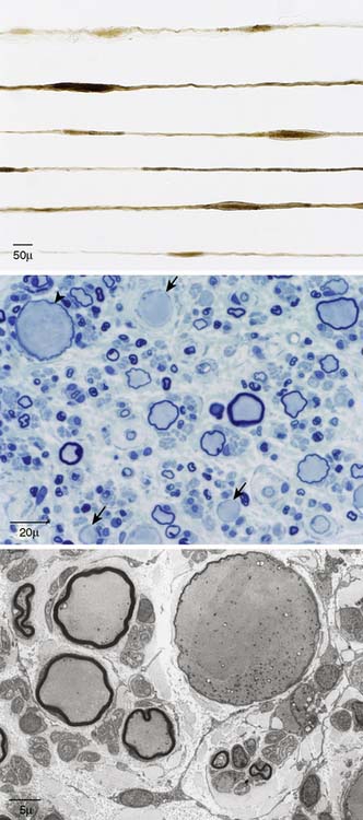
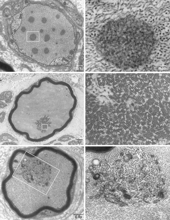
A sural nerve biopsy was performed. At surgery, an enlarged nerve was observed, which finding was supported by morphometric assessment of the transverse fascicular area of 1.24 mm2 (a control nerve of the same age was 0.39 mm2). Teased fiber preparation, paraffin, and epoxy-embedded sections revealed distinctly large axonal swellings (Fig. 28-2). In teased or in longitudinal sections, these enlargements were torpedo shaped and were composed of closely packed intermediate filaments (i.e., neurofilaments). A striking and typical feature was the segregation of intermediate filaments from regions with other axonal elements (i.e., microtubules, mitochondria, smooth endoplasmic reticulum, and dense bodies). In the axonal enlargement were aggregates of closely packed or misdirected neurofilaments (intermediate filament aggregates), and in other cases, the aggregates were composed of non-neurofilament elements (Fig. 28-3).

Figure 28-2 Upper Frame, Different teased nerve fibers of sural nerve showing the torpedo-shaped enlargements along their length. Middle Frame, A transverse semithin epoxy section stained with methylene blue, showing lack (arrows) or thin myelin (arrowhead) of giant axons (spheroids) and, in the lower frame, a low-power electron micrograph of a giant axon and sural myelinated fibers with thin myelin.

Figure 28-3 Different ultrastructural features of transverse sections of spheroids near points of maximal enlargement. Note absence of myelin (left upper frame) and thin myelin relative to axonal caliber of left middle and lower frames. For all low-power electron micrographs (left frame), the majority of the axon is filled by densely packed intermediate filaments (neurofilaments) and typically other elements are sequestered from the neurofilaments, as shown by the broken lines (left upper frame). Within the dense fields of neurofilaments are aggregates of neurofilaments forming denser bodies (right upper and middle frame). These appear as multiple arrays of a few to many neurofilaments at right angles to the direction of axons (right middle frame). A second type of aggregate consists of segregated mitochondria, vesicles and microtubules (right lower frame).
Stay updated, free articles. Join our Telegram channel

Full access? Get Clinical Tree


