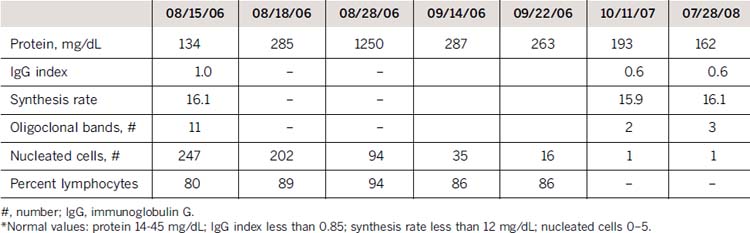
Multiple cerebrospinal fluid (CSF) analyses showed consistently high protein levels (maximum 1250 mg/dL) and cell counts (maximum 250 white blood cells, 89% lymphocytes) but no malignant cells (Table 5-2).
Magnetic resonance imaging (MRI) showed marked enhancement of cauda equina and thoracolumbar nerve roots, multiple cranial nerves, and leptomeninges (Fig. 5-1).
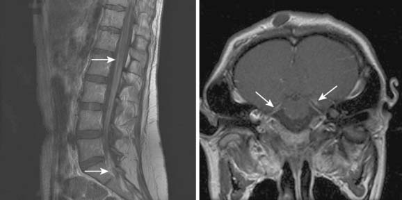
Figure 5-1 Magnetic resonance imaging (MRI) scan of the lumbar spine and cauda equina, showing thickened, gadolinium-enhancing roots (arrows) (left) and brain MRI (right) showing contrast enhancement of cranial nerves VII to VIII bilaterally (arrows).
A fascicular nerve root biopsy was performed. It revealed marked axonal degeneration, numerous macrophages, few T lymphocytes infiltrating endoneurium but neither granulomas nor atypical lymphocytes (Fig. 5-2). Nerve fiber histogram and paraffin sections showed marked fiber loss (Figs. 5-3 and 5-4) and inflammatory infiltrates (see Fig. 5-3). The pattern was believed to be most consistent with a fulminant inflammatory process.
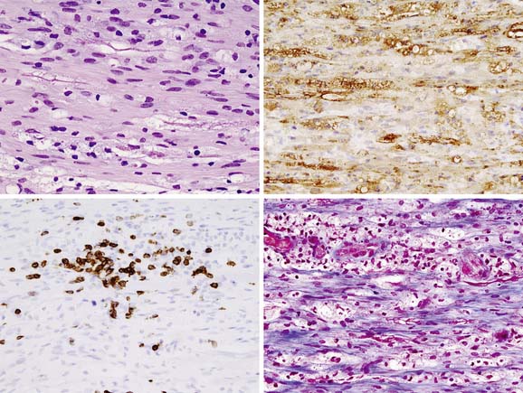
Figure 5-2 The biopsy consisted of spinal nerve demonstrating influx of macrophages (H&E stain, upper left) among nerve fibers with accompanying myelin loss. Loss of uniformity and bubbling of Schwann sheaths was seen (S100 protein immunostain, upper right). Influx of cytologically benign, small T lymphocytes (CD3 stain, lower left), as well as interstitial fibrosis (trichrome stain, lower right) were also evident. Magnification ×400.
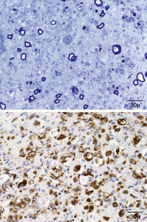
Figure 5-3 Paraffin section of the nerve showing marked fiber loss (upper panel) and CD 68 stain (lower panel) showing the inflammatory infiltrates.
Related posts:
 Malignant Peripheral Nerve Sheath Tumor
Disseminated Sporotrichosis with Multiple Granulomatous Mononeuropathies
Late Sporadic CMT4C—A New KIAA1985 Mutation
Late-Onset Transthyretin Val30Met Familial Amyloid Polyneuropathy Unrelated to Endemic Foci
A Weak, and Numb Patient with Tremor—Antimyelin-Associated Glycoprotein Polyneuropathy
Length-Related Axonal Loss in Neuropathy
Malignant Peripheral Nerve Sheath Tumor
Disseminated Sporotrichosis with Multiple Granulomatous Mononeuropathies
Late Sporadic CMT4C—A New KIAA1985 Mutation
Late-Onset Transthyretin Val30Met Familial Amyloid Polyneuropathy Unrelated to Endemic Foci
A Weak, and Numb Patient with Tremor—Antimyelin-Associated Glycoprotein Polyneuropathy
Length-Related Axonal Loss in Neuropathy
Stay updated, free articles. Join our Telegram channel

Full access? Get Clinical Tree


