Amyotrophic Lateral Sclerosis and its Variants

27 Amyotrophic Lateral Sclerosis and its Variants


Electrodiagnostic (EDX) studies play a central role in the evaluation of patients with amyotrophic lateral sclerosis (ALS), the most common of all motor neuron disorders. Although described earlier by others, the French neurologist Jean-Martin Charcot is credited as naming the disorder amyotrophic lateral sclerosis in 1869. The name is derived from the Greek amyotrophic, which means “no nourishment to the muscle,” lateral, which refers to the lateral area in the spinal cord where the corticospinal tract is located, and sclerosis, which describes the scarring in the spinal cord that occurs when motor neurons deteriorate. In the United States, ALS is commonly referred to as Lou Gehrig’s disease, after the famous baseball player who died of the condition in 1941.


ALS is most often encountered as a sporadic, progressive, degenerative disorder of unknown etiology that characteristically affects both upper motor neurons (UMNs) and lower motor neurons (LMNs) and spares sensory and autonomic function. A small number of cases of ALS (approximately 10%) are familial and are discussed in Chapter 28. In addition, several variants of ALS are well recognized, including progressive bulbar palsy, progressive muscular atrophy (PMA), and primary lateral sclerosis (PLS). Other less common motor neuron disorders exist, including those with atypical motor neuron manifestations caused by genetic mutations, infections, and immunologic disorders (see Chapter 28). Because the prognosis in ALS is uniformly poor compared with other motor neuron disorders, it is essential that the correct diagnosis be reached.


Electromyography (EMG) and nerve conduction studies are most often used to support the diagnosis of ALS. More importantly, however, they are used to help exclude other conditions, some potentially treatable, which may mimic ALS.


Nowhere else is the clinical–electrophysiologic correlation more important than in ALS. EDX studies, by themselves, cannot make a diagnosis of ALS. Rather, ALS remains a clinical diagnosis supported by EDX findings. The electromyographer must appreciate that other disorders may display EDX findings similar to those found in ALS (e.g., coexistent cervical and lumbar radiculopathy) and that it is the combination of clinical and EDX findings that allows a final diagnosis to be reached.


Clinical


Classic Amyotrophic Lateral Sclerosis


ALS is a degenerative, progressive disorder that affects both UMNs and LMNs. Although younger patients may be affected, it occurs most frequently in those 55 to 60 years old, with a slight male predominance. Signs and symptoms of LMN dysfunction include muscle atrophy, weakness, fasciculations, and cramps. UMN dysfunction manifests as stiffness, slowness of movement, spasticity, weakness, pathologic hyperreflexia, and Babinski responses. The presence of both UMN and LMN signs in the same myotome is characteristic of ALS. The mean duration of illness from symptom onset to death is approximately 3 years. However, it is important to remember that about 10% of patients follow a more benign course, surviving for many more years.


ALS is remarkably specific for the motor system. Although detailed pathologic studies have shown some minor loss of sensory fibers, it is distinctly unusual to see sensory complaints or findings on examination. Likewise, there is no disturbance of vision, hearing or the autonomic system. Late in the course, spasticity can affect the bladder, creating symptoms of urinary urgency and frequency. Clinically, an association between abnormalities of cognition and ALS has been recognized in some patients, especially between ALS and Frontotemporal Dementia (FTD). This association is seen in both sporadic and familial forms of ALS and FTD. If patients with classic ALS undergo formal neuropsychological testing, some 40–50% will display some mild evidence of executive dysfunction. FTD develops in approximately 5 to 15% of patients with ALS, and conversely, 10–15% of FTD patients show an associated motor neuron syndrome. Most often, ALS is a regional disease that usually starts in one body segment and progresses to adjacent myotomes. Most cases begin with insidious weakness in either a distal upper or lower extremity. In the upper extremity, the initial presentation can mimic an ulnar neuropathy, especially one at the wrist. In the lower extremity, the presentation is often a progressive foot drop, sometimes misdiagnosed as a peroneal palsy or L5 radiculopathy. As time progresses, symptoms develop in adjacent myotomes of the same limb and then spread to the contralateral limb. Progression continues to other extremities and ultimately to bulbar and respiratory muscles. Death usually results from respiratory insufficiency or from medical complications of prolonged inactivity (pulmonary embolus, sepsis, pneumonia, etc.).


The El Escorial criteria are most often quoted in reaching a diagnosis of ALS. These criteria were set by the World Federation of Neurology meeting in El Escorial, Spain, and published in 1994. They identify four separate body part regions: craniobulbar, cervical, thoracic and lumbosacral. Definite ALS requires that both UMN and LMN signs be seen together in at least three of these regions. Probable ALS requires UMN and LMN signs in two regions, with some UMN signs rostral to the LMN signs. Possible ALS requires UMN and LMN signs in one region or UMN signs in at least two regions. In addition to these criteria, there must be an absence of EDX, pathologic, or radiologic evidence that would support the diagnosis of another disease that may mimic ALS.


Patients with a typical ALS presentation including diffuse atrophy, weakness, fasciculations, and spasticity, in the appropriate age group and clinical setting, are relatively easy to identify. However, not all cases are straightforward, especially when patients present early in the illness with signs and symptoms that are anatomically restricted. In addition, several variants within the spectrum of classic ALS can present diagnostic problems (discussed in the following sections).


Progressive Bulbar Palsy


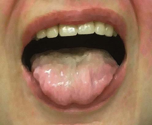
Patients with progressive bulbar palsy initially develop symptoms restricted to the bulbar muscles. They usually present with a several month history of progressive dysarthria with gagging, choking, and weight loss. The speech disturbance may lead to complete anarthria. These patients are commonly incorrectly diagnosed, and many undergo exhaustive ear, nose, and throat or gastrointestinal evaluations looking for the cause of dysarthria or dysphagia. Occasionally, patients may present with respiratory distress as the result of aspiration. Speech is most commonly slow and spastic with variable flaccid features, depending on the degree of LMN dysfunction. The tongue may be atrophied with fasciculations, accompanied by brisk jaw, gag, and facial reflexes (Figure 27–1). One of the characteristic signs is the “napkin or handkerchief sign.” Because of excessive drooling from bulbofacial weakness, patients often carry a tissue in their hand to frequently clear their mouth and face of saliva. Occasionally the symptoms remain relatively restricted to the bulbar muscles. However, in the vast majority of patients the disorder eventually progresses to involve the limbs, as in typical ALS. Indeed, approximately 25% of patients with ALS will have the bulbar onset form.


Differential Diagnosis


The diagnosis of ALS usually is straightforward in patients who present with prominent UMN and LMN signs in both limb and bulbar muscles. However, most patients initially are seen early in the course of the disease, often when only one extremity is clinically affected. In addition, there are other disorders, some potentially treatable, which can mimic the clinical signs, electrophysiologic findings, or both in ALS and its variants (Box 27–1; also see Chapter 28). These disorders are discussed in detail later. In the case of classic ALS, the most important diagnosis to consider is coexistent cervical and lumbar stenosis. For PMA or predominantly LMN presentations of ALS, including the flail arm and flail leg syndromes, the most important diagnoses to consider are demyelinating motor neuropathy, especially MMNCB, and inclusion body myositis (IBM). In addition, benign fasciculation syndrome (BFS) and the myotonic disorders need to be kept in mind. In PLS, there is a large list of neurologic conditions that can be confused with the disorder and need to be excluded by appropriate imaging and other laboratory testing (see the section on Primary Lateral Sclerosis below).


Cervical/Lumbar Stenosis


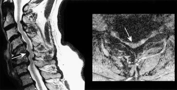
Degenerative disease of the neck and back is extremely common, especially in older individuals. The combination of cervical and lumbar spondylosis occasionally can mimic ALS, both clinically and in the EMG laboratory. Cervical spondylosis, by itself, is a common cause of gait disturbance in the elderly. Compression in the cervical area can result in a polyradiculopathy involving the cervical nerve roots as well as a myelopathy from direct cord compression. This can create a clinical picture of LMN dysfunction in the upper extremities and UMN dysfunction in the lower extremities (Figure 27–2). If additional compression occurs above the C5 level, UMN signs can be seen in the upper extremities as well. To complicate the situation further, patients with coexistent lumbar stenosis may have additional LMN signs in the lumbosacral myotomes. Taken together, the clinical picture can resemble ALS.


However, several points in the history and on the neurologic examination should raise the question of possible cervical or lumbar (or combined) stenosis. Cervical stenosis often follows a stepwise progression, sometimes associated with periods of improvement. In addition, there is usually some neck or radicular pain, along with limitation of neck motion and sensory symptoms in the arms. Paresthesias and vibratory loss in the lower extremities may occur as a result of posterior column compression. A Romberg sign may be present. Back pain commonly accompanies coexistent lumbar stenosis. Moreover, increased pain or sensory disturbance may develop after walking a distance, which is relieved only by the sitting position.


The signs and symptoms noted above will usually suggest the diagnosis of cervical and lumbar stenosis. However, occasionally a patient with cervical and lumbar stenosis presents with a relatively pure motor syndrome consisting of muscle weakness, atrophy, and spasticity, making the clinical distinction from ALS difficult. It is in these patients that the clinical and EMG evaluation of the bulbar and thoracic paraspinal muscles assumes special significance, because they should never be abnormal in lesions restricted to the cervical or lumbar spine (see Chapter 26).


Multifocal Motor Neuropathy with Conduction Block


An important condition that can mimic the PMA presentation of ALS clinically is demyelinating motor neuropathy. Nearly all peripheral neuropathies have both sensory and motor symptoms and signs; therefore, they are not frequently confused with ALS. Very few neuropathies, however, are purely or predominantly motor. Of those, most are demyelinating and are believed to be immune mediated. Although the exact pathophysiology is not understood, presumably some component of motor nerve or myelin is selectively targeted by the immune system, leading to motor dysfunction. It is in these circumstances that a motor neuropathy may be mistaken for a motor neuronopathy (i.e., motor neuron disease). Although it is quite rare, the motor neuropathy that must be excluded, especially in patients with predominantly LMN dysfunction, is MMNCB (see Chapter 26).


MMNCB usually affects only motor fibers, sparing sensory fibers. It often is slowly progressive and begins distally, like ALS. In addition, fasciculations and cramps are common. Unlike ALS, however, it more commonly affects younger patients (<45 years) and has a strong male predominance (male-to-female ratio of approximately 2 : 1). Several important clues may suggest MMNCB on examination. Often, individual motor nerves are affected out of proportion to adjacent nerves that have the same myotomal innervation (hence, multifocal motor neuropathy). For instance, severe weakness in distal median-innervated muscles with relative sparing of ulnar-innervated muscles might occur in MMNCB, but would be very unusual in ALS, marking the disorder as a motor nerve rather than a motor neuron disorder. Second, muscle weakness may appear out of proportion to muscle atrophy in MMNCB, especially early in the course of the disease, reflecting that demyelination, not axonal loss, is the major underlying pathology. Finally, MMNCB does not result in any UMN dysfunction. Reflexes usually are depressed or normal. Pathologic hyperreflexia, spasticity, and Babinski signs are not seen.


The diagnosis of MMNCB may be suggested by the clinical presentation as well as by elevated titers of antiganglioside antibodies, which occur in more than half of patients. Most often, MMNCB is diagnosed through nerve conduction studies, which show evidence of conduction block along motor fibers, between distal and proximal segments. It is extremely important not to miss this diagnosis because the prognosis for these patients is far better than for patients with ALS. Most patients with MMNCB respond well to immune-modulating therapy, especially treatment with intravenous immunoglobulin.


Inclusion Body Myositis


IBM is an idiopathic inflammatory disorder of muscle that can be confused clinically and sometimes electrically with the PMA variant of ALS. IBM is now the most common inflammatory myopathy in individuals older than 50 years. Clinically, IBM presents as slowly progressive weakness. It is more common in men than in women. Along with proximal muscle weakness, distal muscles are commonly involved. In some patients, the distal muscles are weaker than the proximal ones. Although the distribution of weakness most commonly is symmetric, asymmetric presentations often occur. The disease has a predilection for certain muscles, including the iliopsoas, quadriceps, tibialis anterior, biceps, triceps, and long finger flexors. Prominent muscle atrophy, especially of the quadriceps, is common. Facial and ocular weakness does not occur. However, dysphagia is common. The deep tendon reflexes tend to be depressed or absent early in the course, especially the quadriceps reflex. Patients with IBM and severe distal and proximal weakness and wasting, with depressed reflexes, can easily be mistaken for an LMN disease such as PMA.


Unfortunately, the electrophysiology often complicates the diagnosis of IBM. Prominent fibrillation potentials and positive sharp waves are common. Motor unit action potentials (MUAPs) can be small and short, typical of a myopathy; large and long, suggestive of a neuropathic process; or a combination of both. Although large, long duration MUAPs are classically associated with neuropathic disorders, they are also seen in chronic myopathies, especially in those associated with denervation (i.e., usually myopathies with inflammatory or necrotic features).


One of the key differentiating features between LMN disease and IBM is the presence of fasciculations and cramps. Both fasciculations and cramps are neuropathic phenomena; they are not seen in any myopathy, including IBM. In the absence of fasciculations and cramps in a patient with a LMN syndrome, muscle biopsy sometimes is needed to make the differentiation between a motor neuron disorder and IBM.


Stay updated, free articles. Join our Telegram channel

Aug 31, 2016 | Posted by in NEUROLOGY | Comments Off on Amyotrophic Lateral Sclerosis and its Variants

Full access? Get Clinical Tree

Get Clinical Tree app for offline access