Chapter 2 An overview of the brain and pharmacological principles
The information in this chapter will assist you to:
Introduction
In order to understand how psychotropic drugs bring about changes in affect, perception, cognition and/or behaviour an introduction to the way in which the brain is organised and processes information is required. The information in this chapter will provide a reference point for the discussions of pathophysiological processes and the properties of important drug groups that follow in later chapters.
Organisation of the brain
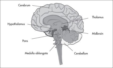
The four principal parts of the brain are the cerebrum, diencephalon, brain stem and cerebellum (see Fig 2.1).

Figure 2.1 The principal parts of the brain.
Adapted from Fundamentals of Pharmacology (5th edn), 2007, by S Bullock, E Manias and A Galbraith, Prentice Hall, Fig 33.1, p 351, with permission
Cerebrum
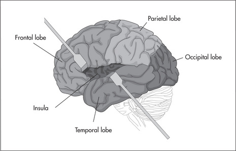
The cerebrum is the largest part of the human brain and its surface is rippled with shallow grooves and ridges that afford the characteristic walnut-like appearance – these undulations increase the surface area of the cerebrum to provide greater processing power. The surface crust of the cerebrum is about 3 mm thick and is called the cerebral cortex. A deep midline groove separates the cerebrum into equal halves called cerebral hemispheres. Communication between hemispheres is achieved via a number of commissures or connecting bridges of nerve fibres, the biggest of these being the corpus callosum. The hemispheres can be further subdivided into lobes, of which there are four external and one internal. The four external are called the frontal, parietal, temporal and occipital lobes; the internal one, tucked in deep between the frontal and temporal lobes, is called the insula (see Fig 2.2).

Figure 2.2 Lobes of the human brain.
Adapted from Human Anatomy and Physiology (7th edn), 2007, by E N Marieb and K Hoehn, Pearson-Benjamin Cummings, Fig 12.6, p 435, with permission
The cerebral cortex is organised in such a way as to allow specialisation of function. Discrete regions of the cortex control the primary processing of vision, taste, hearing, general sensations and muscle movements. Furthermore, as new information is received it can be referenced against past experiences stored in association areas. The frontal lobes are particularly important in the processing of emotions and behaviour, decision making, personality and temperament, self-discipline and cognition. Another form of specialisation occurs at the hemispheric level: one hemisphere may be more dominant over the other in the control of some functions. Classic examples of this cerebral dominance include handedness (90% of humans are right-handed), language control (left hemisphere is usually dominant) and spatial perception (right hemisphere is usually dominant). This phenomenon does not mean that the non-dominant hemisphere makes no contribution to these functions, nor does it mean that in a typical right-handed person their left hemisphere is dominant for all of these functions – it is very much function-specific. In some brain disorders, such as schizophrenia and depression, an imbalance between the hemispheres may contribute to the manifestation of the condition (see Chs 3 and 4).
Brain stem and cerebellum
The cerebellum has a major role in muscle movement. It assists in the planning of movements, provides feedback to the cortex as to the efficiency of the initiated movements, is involved in the control of posture and muscle tone and ensures that movements are smooth and coordinated.
Neuronal communication
Neurotransmission
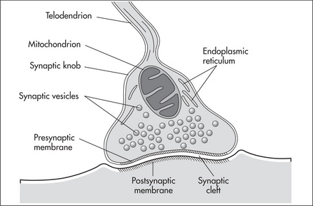
The interface or connection between sending and receiving neurones is called the synapse. It has been suggested that there are 1 × 10¹⁵ synapses in the human brain. The synapse consists of the axon terminal of the sending neurone (the presynaptic neurone), the cell membrane of the receiving neurone (the postsynaptic neurone) and a small gap between the two called the synaptic cleft (see Fig 2.3). As the presynaptic and postsynaptic neurones do not actually make direct contact, the communication is conveyed postsynaptically via the release of chemical messengers or neurotransmitters.

Figure 2.3 The synapse.
A representation of a typical synapse.
Reproduced from Fundamentals of Anatomy and Physiology (7th edn), 2006, by F H Martini, Pearson Education, Inc (publishing as Benjamin Cummings), Fig 12.2, with permission
Stay updated, free articles. Join our Telegram channel

Full access? Get Clinical Tree


