Chapter 1
Anatomy of the Larynx
The larynx develops from the endodermal lining and the mesoderm of the fourth to sixth branchial arches. At birth the tip of the epiglottis is at the level of the first cervical vertebra and may come in contact with the soft palate. Up to the age of 3 years the larynx descends in the neck and then little change occurs until puberty.1 The larynx in the child is found between cervical vertebra three and five. At puberty the cricoid cartilage descends to the level of the sixth cervical vertebra, which results in part from growth of the thyroid cartilage. The latter cartilage increases in size and shape, especially in males. Growth of the thyroid prominence causes the angle between the thyroid laminae to become more acute in the male than in the female, hence the characteristic “Adam’s apple.” The adult male larynx is larger than that of the female.
The laryngeal framework comprises cartilages, muscles, membranes, and ligaments. It is lined with mucosa both within its cavity and on its pharyngeal surface. The mucosa of the cavity is continuous superiorly with the pharynx and inferiorly with that of the trachea. The mucosa of the outer surface of the larynx is the mucosa of the laryngopharynx. Contraction of the muscles will alter the positions of the cartilages and ligaments during respiration, phonation, and deglutition.
Laryngeal Framework
The larynx comprises nine cartilages—three unpaired cartilages and three sets of paired cartilages. The unpaired cartilages are the thyroid, cricoid, and epiglottis. The paired cartilages, the arytenoids, are of more importance than the corniculates and cuneiforms.
Even though the hyoid bone is technically not part of the larynx, it is involved in movement of the larynx and therefore needs to be discussed with the larynx. The hyoid bone is a U-shaped bone consisting of a body and greater and lesser horns on each side. It derives from mesoderm of the second and third branchial arches and serves as attachment for numerous muscles of the tongue as well as some of the extrinsic muscles of the larynx. It is suspended from the tip of the styloid process of the temporal bone by the stylohyoid ligament and attached to the larynx by the thyrohyoid membrane. Because muscles of the tongue and larynx are attached to it, the hyoid bone plays an important role in movement of both structures.
Cartilages
Thyroid Cartilage (Figs. 1.1 and 1.2)
The thyroid cartilage, the largest of the laryngeal cartilages, comprises two quadrilateral laminae fused in the midline as the laryngeal prominence. The angle formed at the prominence is more acute in the male adult (90 degrees) than the female (120 degrees). The superior border of the laminae meet in the midline in a V-shaped notch. Posteriorly, the laminae are incomplete and their edges become elongated as the inferior and superior horns. This configuration leaves the posterior aspect of the larynx open, with the laryngopharynx completed by the pharyngeal musculature. On the external surface of the laminae is the oblique line, a ridge that extends from the superior thyroid tubercle located inferior to the superior horn on its posterolateral edge. It extends inferomedially to the inferior thyroid tubercle. Three muscles attach to the oblique line (sternothyroid, thyrohyoid, and inferior constrictor). The superior border of the thyroid cartilage is concave and gives attachment to the thyrohyoid membrane. The superior horn is the lateral-most edge of attachment of the membrane, which is thickened in this area. This is the lateral thyroid ligament. The inferior border is concave posteriorly and convex anteriorly and gives attachment to the cricothyroid ligament. The inferior horn articulates with the cricoid cartilage at its lower end, forming the synovial cricothyroid joint. The thyroid cartilage comprises hyaline cartilage, which ossifies with age. Ossification usually begins at approximately 25 years of age.
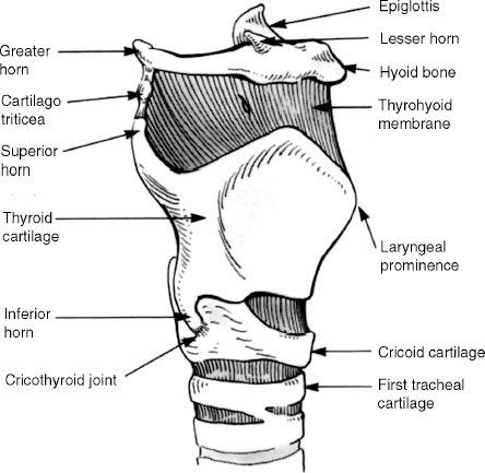
Fig. 1.1 Oblique view of the larynx.
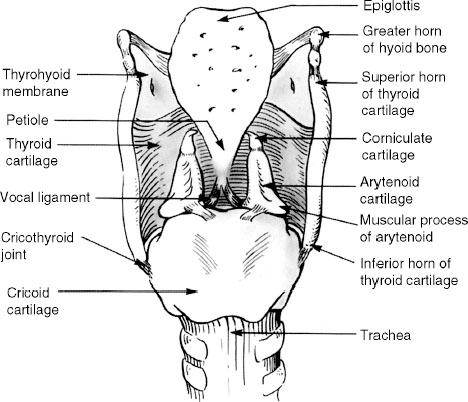
Fig. 1.2 Posterior view of the larynx.
Cricoid Cartilage (Figs. 1.1 and 1.2)
The cricoid cartilage lies between the thyroid cartilage and the trachea. In contrast to the thyroid cartilage, it is a complete ring, “signet” in shape. Posteriorly it has a large quadrate-shaped lamina that narrows into the arch anteriorly. The posterior lamina, which is 2 to 3 cm in vertical height, has a median vertical ridge with shallow depressions on each side. The posterior cricoarytenoid muscles originate in these depressions. The superior border of the lamina has two small oval convex facets that serve as the articular surfaces for the arytenoid cartilages. The cricoid’s articular facet for the cricothyroid joint is at the point where the lamina narrows to become the arch. The cricoid cartilage is hyaline cartilage and with age undergoes various amounts of ossification.
Arytenoid Cartilage (Figs. 1.2, 1.3, and 1.4)
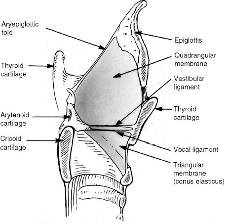
Fig. 1.3 Midsagittal cut of the larynx showing fibroelastic membrane attachments.
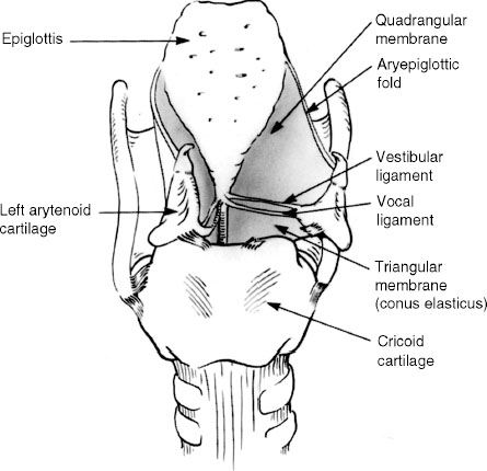
Fig. 1.4 Posterior view of the larynx showing fibroelastic membrane attachments. Arytenoid cartilage on right moved laterally to demonstrate membrane attachments to the ligaments.
Corniculate and Cuneiform Cartilages (Fig. 1.2)
The corniculate cartilages, comprising elastic cartilage, are paired and sit on the apex of the arytenoid cartilages. They extend the apex posteromedially. A synovial joint is usually formed with the arytenoid cartilage, but fusion can occur. The cuneiform cartilages are small, paired, elongated elastic cartilages that are located in the aryepiglottic fold. A small cartilage, the triticeal cartilage, also elastic in nature, is frequently found in each lateral thyrohyoid membrane.
Epiglottis (Figs. 1.2, 1.3, and 1.4)
Related posts:

Stay updated, free articles. Join our Telegram channel

Full access? Get Clinical Tree


