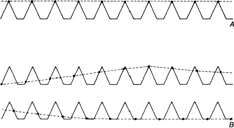
Chapter 10 Stroboscopic examination of the larynx is one of the minimum essential procedures of modern laryngologic practice. The key event in voice production is vibration of the vocal folds. The vibratory behavior of the vocal folds is one of the most important and crucial determinants of the voice character. Abnormal voices are always associated with abnormal vibratory patterns of the vocal fold. Examination of vocal fold vibration, therefore, is necessary and essential to determine the cause and mechanism of abnormal voices. Stroboscopy is the only existing modality to observe vocal fold vibration that is available for clinical purposes. Stroboscopic light sources produce intermittent flashes of light that are synchronous with the vibratory cycles of the vocal folds. The waveform of the patient’s voice picked up with a microphone triggers the light source. When the frequency of the light flashes emitted is the same as that of the vocal fold vibration, a clear still image of the vocal folds at a given phase point is observed (Fig. 10.1A). When the frequency of the flashes is slightly less than that of the vocal fold vibration, resulting in a systematic phase delay of the consecutive light flashes, a slow motion effect is obtained (Fig. 10.1B). Stroboscopy cannot demonstrate fine details of each individual vibratory cycle, but it depicts a vibratory mode averaged over many successive vibratory cycles. If successive vibrations are completely periodic and the vibratory behavior is perfectly uniform, the stroboscopic images reflect precisely the slow-motion images of each vibratory cycle. In humans, however, successive vibrations are aperiodic to a greater or lesser extent. Nevertheless, it is extremely useful for clinical purposes. Fig. 10.1 Schematic presentation of the principle of stroboscopy. (From Hirano M. Clinical Examination of Voice. Vienna—New York: Springer Verlag; 1981. Reprinted with permission.) Figure 10.2 schematically depicts normal vibratory pattern of the vocal fold in modal register and Fig. 10.3 shows vibratory phases in one vibratory cycle. The vibratory cycle consists of three phases: the opening phase, in which the vocal fold edges move laterally; the closing phase, in which the vocal edges move medially; and the closed phase, in which the bilateral vocal folds are in contact with each other. Typically, two wave peaks are observed on the vocal folds. They are referred to as the upper and lower lips. The upper and lower lips are not structures that are located at consistent places of the vocal fold, but they are the peaks of waves that travel on the vocal fold mucosa. At the end of the closed phase, the vocal folds are in contact with each other only at the upper lip (Fig. 10.2A). During the early stage of the opening phase, the entire vocal fold shifts laterally (Fig. 10.2B). At the maximum opening the upper and lower lips are lined up on the same plane (Fig. 10.2C). In the early stage of the closing phase, the lower lips move medially whereas the upper lips still move laterally (Fig. 10.2D)
Stroboscopic Examination of the Normal Larynx
How Stroboscopic Images of Vocal Fold Vibrations Are Obtained
Normal Vibratory Pattern of Vocal Folds
Related posts:
![]()
Stay updated, free articles. Join our Telegram channel


Full access? Get Clinical Tree


